6. Conclusions and Future Directions
The developmental program of bacterial cell division ensures divisions take place with high temporal and spatial fidelity. A molecular machine, called the divisome, executes this process. The underlying molecular mechanism of this process has historically been studied from a genetic and cell-biological point of view. That is, large amounts of data have been accumulated regarding the genes involved in cell division, their expression profiles and final localization of gene products during the cell cycle by EM, immunofluorescent and GFP-fusion technologies. Genetically modified strains have also been utilized to examine the effects of mutations and depletion of various components in the divisome, largely by mapping the stability and localization of the remaining components. These results have lead to a number of hypotheses regarding protein-protein interactions in the divisome and the construction of a hierarchical model for the assembly of the divisome (see Figure 1.4). While this model holds well for E. coli, there appears to be more cooperativity in the assembly of the divisome in B. subtilis (discussed in Section 1.2.3). Regardless, this map has begun to describe how the divisome assembles.
The proteins, DivIB, FtsL and DivIC, appear to cooperatively assemble to the B. subtilis divisome, although other proteins are likely to be also required for assembly of this group (see Section 1.3.2). While depletion and localization studies have established this cooperativity, there has been no attempt to explain mechanistically how they come to coalesce. What makes these proteins even more puzzling is their primary structures have not aided in understanding their role either.
The results presented in chapter three of this thesis established for the first time, that the extracytoplasmic domains of FtsL and DivIC were intrinsically unstable and do not interact with themselves or one another, despite having apparent coiled-coil motifs. It was also shown that addition of the extracytoplasmic domain of DivIB is not sufficient to generate a ternary complex either. Recently, however, two reports have indicated that DivIB, FtsL and DivIC can form a ternary complex when placed in an appropriate context. Most likely, this context models insertion of FtsL and DivIC into membranes (discussed in Section 3.4). While the establishment of complex formation of these three proteins is, in some ways, gratifying, currently there is still no meaningful interpretation for the role of these proteins in the divisome. An analysis of the structure of this complex at the atomic level could aide in this interpretation.
The extracytoplasmic region of DivIB/FtsQ is necessary and sufficient for its divisomal localization and cell division function (Chen, Weiss et al. 1999; Katis and Wake 1999). However, prior to this study, nothing was known about its molecular architecture. In this study, it was demonstrated that the extracytoplasmic region of DivIB comprises three domains, designated , , and from N- to C-terminus. The and domains are structurally autonomous whereas the domain is proteolytically sensitive and presumably unstructured in the absence of other divisomal proteins. The entire domain architecture of DivIB was defined as shown in Figure 4.2B, and the architecture of the entire DivIB/FtsQ family is expected to be very similar based on sequence homology with DivIB.
The domain was almost exactly coincident with the polypeptide-transport-associated (POTRA) domain that was predicted on the basis of bioinformatic analyses to be present in DivIB/FtsQ and in a class of -barrel outer-membrane proteins involved transport or assembly of polypeptides (Sanchez-Pulido, Devos et al. 2003). The POTRA domain has been hypothesized to act as a chaperone for unfolded peptides (discussed in Section 4.6). Does the domain of DivIB act as an intramolecular chaperone for the domain? Given that the domain is readily degraded by proteases in the presence of the domain (Figure 4.1A) this seems unlikely. Instead, one possible role the domain may have is in binding and stabilizing FtsL and/or DivIC which, as described above, are otherwise unfolded in the absence of other divisomal proteins. Indeed, it is entirely possible that the domain is, at least in part, responsible for stabilizing the ternary interaction between DivIB, FtsL and DivIC, although other regions of DivIB cannot be ruled out as playing a role as well. This hypothesis could be tested in the system reported for S. pneumoniae by Noirclerc-Savoye and co-workers (Noirclerc-Savoye, Le Gouellec et al. 2005) by attempting their pulldown experiments in the absence of the domain. Alternatively, an attempt to complement a divIB null strain by mutant genes for divIB in which the domain has been replaced by an irrelevant yet similar sized domain could be attempted.
Despite extensive mutagenesis of the conserved surfaces of the domain (Chapter 5), no part of these surfaces appears to be critical for DivIB function. Indeed, the role of the domain remains enigmatic. Experiments in which divIB genes with the domain swapped with irrelevant domains of similar size are used to complement a divIB null strain could be attempted to see if this domain is critical at all for DivIB function. Notwithstanding, the 3-dimensional structure of the domain is unique and as such is a potential target for drug development.
Remarkably, the domain, despite being relatively small in size (~30 residues) and unstructured, is absolutely critical for function (see section 5.2.4). Furthermore, it has been shown that deletion of the C-terminal 29–30 residues of FtsQ from E. coli, which corresponds to the bulk of the domain – as mapped in this thesis – abrogates its interaction with all other divisomal proteins in bacterial two-hybrid assays (Karimova, Dautin et al. 2005). In addition, mutations in the domain of FtsQ fail to recruit FtsL and other downstream divisomal components in vivo (Chen, Minev et al. 2002). Thus, is not essential for DivIB/FtsQ localization, however, it appears to be the most critical domain for mediating the interaction of DivIB/FtsQ with other components of the divisome.
The extracytoplasmic regions of FtsL and DivIC are predicted to comprise a membrane-proximal coiled coil region of ~35 residues followed by a 25–30 residue C-terminal region of unknown structure. As for DivIB, all reported mutant alleles of FtsL and DivIC map to the C-terminal domain (Levin and Losick 1994; Sievers and Errington 2000). In striking contrast, the coiled coil region of B. subtilis FtsL can be extensively mutagenized and replaced with heterologous coiled coils with as little as 34% sequence identity without affecting cell division (Sievers and Errington 2000).
Taken together, these data suggest that the interaction between FtsL, DivIC, and DivIB is most likely mediated by their C-terminal regions. Such an interaction would be difficult if the domain of DivIB adopted the trans conformation. In this conformation, the N- and C-termini of the domain are located in close proximity and the domain is directed towards the membrane (Figure 6.1, left panel). If the domain is directed towards the membrane, how can it make cognate interactions with the C-terminal regions of FtsL and DivIC?
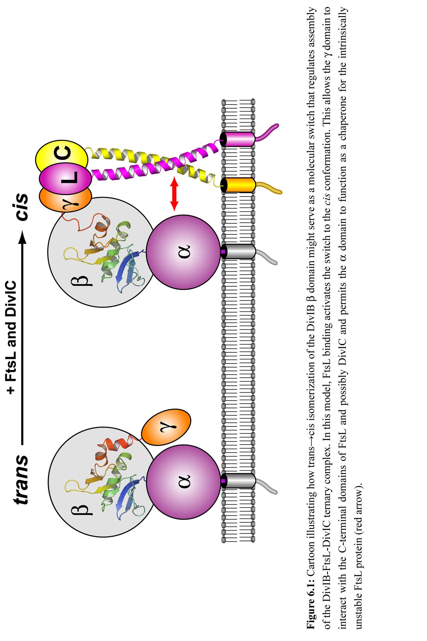
One possibility is that cis-trans isomerization of the domain serves as a molecular switch that modulates assembly of the FtsL-DivIC-DivIB complex. Interaction of DivIB with FtsL might activate this switch, causing the domain to isomerize to the cis conformer. In this conformation (Figure 6.1, right panel), the domain is located distal to the membrane and suitably positioned for interaction with the C-terminal domains of FtsL and DivIC. In addition to orienting the domain for interaction with FtsL and/or DivIC, trans→cis isomerization might position the domain so it can serve as a chaperone for FtsL and in addition help stabilize an FtsL/DivIC interaction via their coiled-coil domains. Thus, a cis-trans switch might serve two purposes: regulation of the interaction of with other divisomal components, and modulation of the chaperone activity of . Although the cis-trans switch might operate in vivo, it is not essential for vegetative cell division as it is possible to rescue a B. subtilis divIB null at the nonpermissive temperature by ectopic expression of divIB containing a P225A mutation (equivalent to P222A in Gste-DivIB), which is expected to abrogate isomerization (Figure 5.3). Similarly, an E. coli ftsQ null strain can be complemented by a multicopy
plasmid expressing an ftsQ(Y227P228AA) double mutant (N. Goehring, personal communication). However, these experiments do not exclude the possibility that cis-trans isomerization acts as a molecular switch that increases the efficiency of septation or that it plays a role in polar divisions during sporulation in B. subtilis.
Given that DivIB, FtsL and DivIC form a complex, and most likely do so by their extreme C-terminal regions (perhaps aided in part by the domain acting as a chaperone) it still remains an open question as to what this complex actually achieves during division. As discussed in Section 1.3.3, this complex may be a regulation step in division in which stability of FtsL must be maintained to complete division. To answer this question, the mechanism of how this complex interacts with members of the divisome that assemble after it will need to be examined. Alternatively, the complex of these three C-terminal regions may form an apparatus with a more direct role in cell division. For example, supply some necessary enzymatic activity or membrane manipulation that permits completion of division. Either way, if the domain of DivIB were responsible for interactions with FtsL and/or DivIC, structural determination of this complex would bring several insights into how this complex forms and aid in the development of antibiotics that target this crucial interaction.