5. Mutational Analysis of DivIB
The extracytoplasmic domain of DivIB/FtsQ contains the necessary and sufficient function(s) of the protein to execute its role in bacterial cell division. In addition, DivIB appears capable of forming a complex with FtsL and DivIC and it seems likely that at least one role of DivIB is to protect FtsL from degradation; this in turn allows proper formation of the divisome and execution of the division event. Prior to the results presented in chapter 4 of this thesis, there was no structural information regarding this crucial region.
As established in chapter 4, DivIB contains three domains, with the domain containing an entirely novel fold. Furthermore, bioinformatic analysis, to date, has not identified homologous sequences for the domain in any other protein (as apposed to the domain). As such, it would appear that the structure of the domain presented in chapter 4 might well be unique to the DivIB/FtsQ family of proteins with no other representatives elsewhere in life. These properties of necessity and uniqueness, along with the cell surface or “exposed” position of the extracytoplasmic domain, at least in Gram positive bacteria, make DivIB an attractive target for rational drug design and anti-microbial development.
An important step in rational drug design is the discovery of critical surface residues for a proteins function. In order to elucidate the important amino acid residues for protein-protein interactions between DivIB and potential binding partners, alanine mutagenesis of single amino acids within DivIB was employed within an in vivo system. This chapter presents the rationale behind the mutants generated and the results of these mutations.
5.1 Determination of surface exposed, conserved residues.
Figure 5.1 presents an alignment of the primary structure of Gste-ecDivIB with the extracytoplasmic regions from other Gram positive bacteria as well as the periplasmic region of FtsQ from the Gram negative bacteria, E. coli. Several of these species are pathogenic to humans, including B. anthracis (anthrax), S. pneumoniae and E. coli itself. The secondary structure elements for the domain (predicted) and domain (determined in chapter 4) are illustrated above the sequence alignment. Buried hydrophobic residues in Gste-DivIB are marked with green triangles below the alignment. Identities are boxed in yellow and conservative substitutions colored orange. This alignment aides in the identification of surface exposed residues that are conserved across species.
5.1.2 The DivIB/FtsQ family is likely to have a consistent protein fold
In the sequence alignment in figure 5.1, the buried hydrophobic residues, (as judged from the Gste-DivIB structure are marked with green triangles (30 in total). Of these, 24 are conserved at greater than 85% consistency. This high level of consistency suggests that the buried core of DivIB/FtsQ is also highly conserved and likely to give rise to a very similar folded structure across the family. In addition, mutation of these conserved hydrophobic residues would have: (i) little direct effect on the ability of DivIB to bind other proteins and; (ii) interfering with the hydrophobic core of the protein and could
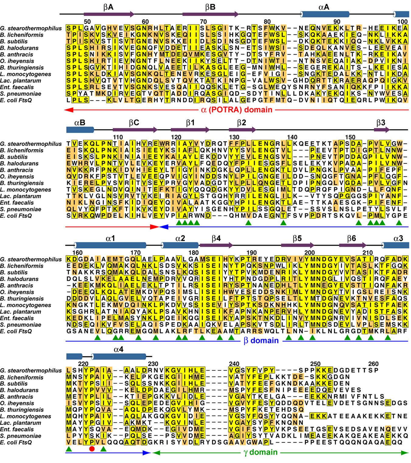
Figure 5.1: Alignment of the primary structure of Gste-ecDivIB with sequences of the extracytoplasmic regions of DivIB from other Gram positive bacteria as well as the periplasmic region of the orthologous FtsQ from Gram negative E. coli. The numbering refers to Gste-ecDivIB. The secondary structure of the (POTRA) domain is a prediction based on multiple sequence alignments , whereas the secondary structure of the domain was experimentally determined in chapter 4. Sequences were aligned using ClustalW . Identities are boxed in yellow, and conservative substitutions are shaded orange. The proline that undergoes cis-trans isomerization in the isolated domain is marked with a red circle. Buried hydrophobic residues in the Gste-DivIB structure are highlighted with green arrowheads. The genus abbreviations refer to Geobacillus (G), Bacillus (B), Oceanobacillus (O), Listeria (L), Lactobacillus (Lac.), Enterococcus (Ent.), Streptococcus (S), and Escherichia (E).
potentially generates false positives for inactivity. Thus, these highly conserved residues were avoided when generating mutants.
5.1.3 Selection of conserved surface residues for mutation
Aside from the conserved buried hydrophobic residues discussed above, figure 5.1 shows there are only three areas of high conservation (yellow and orange shading) in the primary structures of DivIB/FtsQ, namely, between residues 130-140, 180-185 and 199-207. Many of these residues are present on the surface of Gste-DivIB. Figure 5.2 is a molecular surface plot of Gste-DivIB with surface conservation shaded in orange (>75%) and red (>85%). Most of the surface is poorly conserved (shaded grey), yet there are two patches of high homology. Surface 1 (Figure 5.2; left structure) is composed of amino acids S183, E184, Y200, N202, D203, G204, Y221 and P222. P189 was additionally considered for mutagenesis as it is reasonably well conserved and, at one point, was considered a potential site for the proline isomerization discussed in chapter 4. Surface 2 (Figure 5.2; right structure) is composed of amino acids E117, W118, L134, E135, N136 and G137. Of these amino acids, all, except W118, G137 and G204 were targeted for alanine mutagenesis. G137 and G204 were not mutated as these glycine residues participate in type I/II turn secondary structure, where, for steric hindrance reasons, glycine residues are usually strictly required. W118 was also avoided as a change from the bulky tryptophan residue to alanine was considered too radical. Thus, 12 residues were targeted for alanine mutagenesis; namely E117, L134, E135, N136, S182, E184, P189, Y200, N202, D203, Y221 and P222 (see also table 5.1). Note that these residue numbers refer to the number for G. stearothermophilus DivIB. As
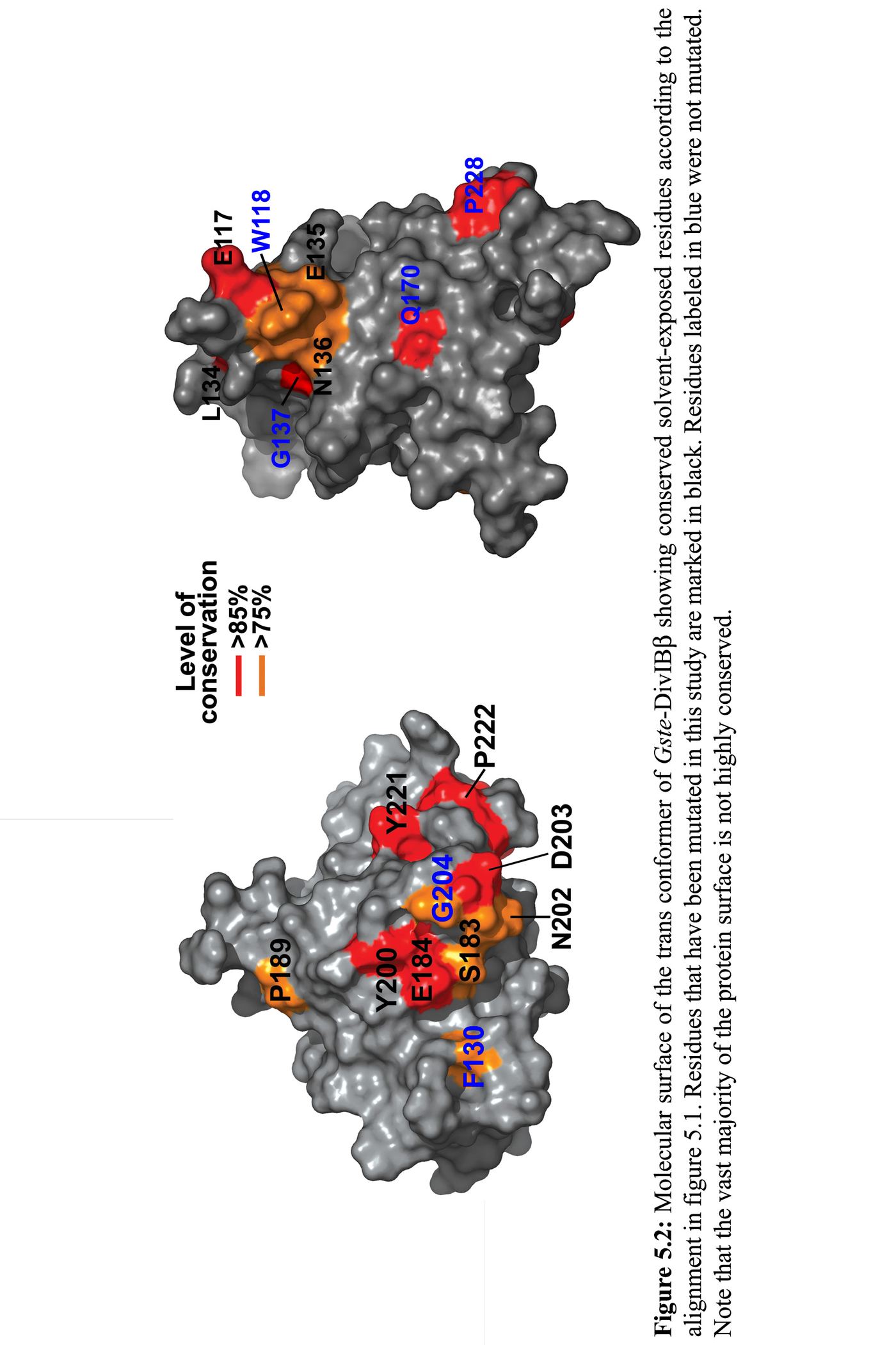
mutational analysis was conducted in an in vivo system in B. subtilis, the equivalent residues in B. subtilis were targeted. Table 5.1 (columns 2 and 3) serves as a key to match residues between B. subtilis and G. stearothermophilus.
5.1.4 Selection of additional residues for mutation
Based on high sequence homology, several residues were selected for mutagenesis in the region proximal to the membrane on the cytoplasmic side (see figure 3.2). These residues are, in B. subtilis, Q26, K27, N29, R30 and R31. Interestingly, the presence of R30 and R31, which is highly conserved across Bacillus species, is in common with many proteins exported across the cytoplasmic membrane by the Tat (twin-arginine translocation) pathway. The Tat pathway was originally identified in chloroplasts (Settles, Yonetani et al. 1997; Santini, Ize et al. 1998) and a similar system appears to operate in E. coli (Weiner, Bilous et al. 1998) and B. subtilis (Pop, Westermann et al. 2003). It was expected that mutations in this region might interfere with the export of and/or membrane localization of DivIB.
Additionally, the region was targeted for mutagenesis as previous mutations have all mapped to the domain (discussed in Section 1.2.2.3). A single well conserved tyrosine (Y246 in B. subtilis) was selected for mutation along with a deletion of the entire domain. The entire list of mutations is summarized in table 5.1.
Table 5.1: List of strains used to test the effect of various point mutations in the divIB gene and resultant phenotypes at 30º C, 42º C and 48º C.
| DomainDomainDomain | GsteGsteGste Residue #Residue #Residue # | BsubBsubBsub MutationMutationMutation | StrainStrainStrain ### | PhenotypePhenotypePhenotype (30º C)(30º C)(30º C) | PhenotypePhenotypePhenotype (42º C)(42º C)(42º C) | PhenotypePhenotypePhenotype (48º C)(48º C)(48º C) |
|---|---|---|---|---|---|---|
| N/AN/AN/A (controls)(controls)(controls) | --- | Wildtype (186)Wildtype (186)Wildtype (186) | SU5SU5SU5 | coloniescoloniescolonies | coloniescoloniescolonies | coloniescoloniescolonies |
| --- | divIB ::catdivIBdivIB :: ::catcat | SU321SU321SU321 | coloniescoloniescolonies | no coloniesno coloniesno colonies | no coloniesno coloniesno colonies | |
| --- | SU321SU321SU321 cat::ermCcatcat::::ermCermC | RSA8RSA8RSA8 | coloniescoloniescolonies | no coloniesno coloniesno colonies | no coloniesno coloniesno colonies | |
| --- | RSA8 amyE::divIB-catRSA8 RSA8 amyEamyE::::divIB-catdivIB-cat | RSA9RSA9RSA9 | coloniescoloniescolonies | coloniescoloniescolonies | coloniescoloniescolonies | |
| CytoplasmicCytoplasmicCytoplasmic | Q21Q21Q21 | Q26AQ26AQ26A | RSA11RSA11RSA11 | coloniescoloniescolonies | not testednot testednot tested | coloniescoloniescolonies |
| K22K22K22 | K27AK27AK27A | RSA12RSA12RSA12 | coloniescoloniescolonies | not testednot testednot tested | coloniescoloniescolonies | |
| N24N24N24 | N29AN29AN29A | RSA13RSA13RSA13 | coloniescoloniescolonies | not testednot testednot tested | coloniescoloniescolonies | |
| R25R25R25 | R30AR30AR30A | RSA14RSA14RSA14 | coloniescoloniescolonies | not testednot testednot tested | coloniescoloniescolonies | |
| R26R26R26 | R31AR31AR31A | RSA15RSA15RSA15 | coloniescoloniescolonies | not testednot testednot tested | coloniescoloniescolonies | |
| E117E117E117 | E122AE122AE122A | RSA24RSA24RSA24 | coloniescoloniescolonies | not testednot testednot tested | coloniescoloniescolonies | |
| L134L134L134 | L139AL139AL139A | RSA16RSA16RSA16 | coloniescoloniescolonies | not testednot testednot tested | coloniescoloniescolonies | |
| E135E135E135 | E140AE140AE140A | RSA17RSA17RSA17 | coloniescoloniescolonies | not testednot testednot tested | coloniescoloniescolonies | |
| N136N136N136 | N141AN141AN141A | RSA18RSA18RSA18 | coloniescoloniescolonies | not testednot testednot tested | coloniescoloniescolonies | |
| S183S183S183 | S186AS186AS186A | RSA25RSA25RSA25 | coloniescoloniescolonies | not testednot testednot tested | coloniescoloniescolonies | |
| E184E184E184 | E187AE187AE187A | RSA26RSA26RSA26 | coloniescoloniescolonies | not testednot testednot tested | coloniescoloniescolonies | |
| P189P189P189 | P192AP192AP192A | RSA27RSA27RSA27 | coloniescoloniescolonies | not testednot testednot tested | coloniescoloniescolonies | |
| Y200Y200Y200 | Y203AY203AY203A | RSA19RSA19RSA19 | coloniescoloniescolonies | not testednot testednot tested | coloniescoloniescolonies | |
| N202N202N202 | N205AN205AN205A | RSA21RSA21RSA21 | coloniescoloniescolonies | not testednot testednot tested | coloniescoloniescolonies | |
| D203D203D203 | D206AD206AD206A | RSA22RSA22RSA22 | coloniescoloniescolonies | not testednot testednot tested | coloniescoloniescolonies | |
| Y221Y221Y221 | Y224AY224AY224A | RSA30RSA30RSA30 | coloniescoloniescolonies | not testednot testednot tested | coloniescoloniescolonies | |
| P222P222P222 | P225AP225AP225A | RSA31RSA31RSA31 | coloniescoloniescolonies | not testednot testednot tested | coloniescoloniescolonies | |
| Y243Y243Y243 | Y246AY246AY246A | RSA28RSA28RSA28 | coloniescoloniescolonies | colonies/colonies/colonies/ early lysisearly lysisearly lysis | no coloniesno coloniesno colonies | |
| deletion deletiondeletion | deletion deletiondeletion 237-262237-262237-262 | RSA29RSA29RSA29 | coloniescoloniescolonies | no coloniesno coloniesno colonies | no colonies§no coloniesno colonies§§ |
5.2 Mutagenesis of surface exposed, conserved residues.
To test the importance of each surface residue discussed above, an in vivo system was developed to test for DivIB function in the presence of mutations. This allowed for rapid screening of mutations and dissection of critical residues.
5.2.1 Construction of experimental system.
The basis of this experimental system exploited the fact that a divIB null strain of B. subtilis will form colonies on an LB plate at 30º C; however, at 48º C, cell division fails and colonies do not form (Beall and Lutkenhaus 1989; Harry and Wake 1989). While complementation and, hence, recovery of this mutation by an ectopic copy of the wildtype divIB gene has not been previously reported, it was expected that a divIB null strain should grow at 48º C by integration of wildtype divIB under its native promoter at the amyE locus in B. subtilis.
In order to produce the necessary strains required to test complementation, the strain SU321 (Harry and Wake 1989) was sourced as a divIB null strain. This strain contains a complete disruption of the divIB gene at its native locus by a chloramphenicol resistance gene. For chromosomal integration of ectopic copies of divIB (and its mutants), derivatives of the pDG364 plasmid, carrying wildtype and mutant copies of the divIB gene, were constrcuted. The pDG364 plasmid also carries a chloramphenicol resistance cassette and permits integration of ectopic genes and chloramphenicol resistance at the amyE locus. Because SU321 was already resistant to chloramphenicol, the antibiotic resistance of SU321 was converted to erythromycin by first transforming SU321 with pCm:Er (Steinmetz and Richter 1994). The resultant strain, RSA8, was verified as resistant to erythromycin, not resistant to chloramphenicol, and displayed the temperature-sensitive phenotype of SU321. Thus, transformation of RSA8 with the pDG364 vector and derivatives of this vector results in strains that are resistance to chloramphenicol and erythromycin. The entire method is described in section 2.x.x.
Testing of complementation by ectopic copies of divIB and mutants of divIB was achieved by simply streaking strains on LB plates, at 30º C and 48º C and checking for colony growth.
5.2.2 Testing of control strains
Table 5.1 summarizes the complementation results for the entire list of mutants described in section 5.1.3 and 5.1.4. The first four rows detail the results for the control strains. Wildtype B. subtilis (SU5, 186) successfully forms colonies at all tested temperatures, namely 30º C, 42º C and 48º C (also see figure 5.4, each plate, sectors between 10 and 12 o’clock). When tested in this study, the divIB null strain (SU321) formed colonies at 30º C and failed to form colonies at 42º C and 48º C. The strain RSA8 (divIB::cat::ermC) also formed colonies at 30º C and failed to form colonies at 42º C and 48º C (also see figure 5.4, each plate, sectors between 12 and 2 o’clock). The RSA9 strain was constructed by transformation of RSA8 with pSAR50 (pDG364 into which the wildtype divIB gene has been inserted, see section 2.x.x.). This placed an ectopic copy of the divIB gene at the amyE locus; thus, this strain was used as a positive control in this complementation study. In table 5.1, it can be seen that RSA9 forms colonies at 30º C, 42º C and 48º C. There are no apparent differences between RSA9 and wildtype B. subtilis under these conditions. The colony forming ability of RSA9 can be seen in figure 5.4 (sectors between 2 o’clock and 4 o’clock).
5.2.3 The cytoplasmic and domain of DivIB is malleable
To test for the ability of mutant divIB genes to complement RSA8, strains were constructed by transforming RSA8 with derivatives of pSAR50 (see section 2.x.x.). These pSAR50 derivatives contained point mutated divIB genes. In all cases where point mutations were generated in the cytoplasmic and domain portion of divIB, the mutated gene complemented fully at 48º C (see table 5.1). Figure 5.3 illustrates the results for two such mutants, the Y224A (strain RSA30) and the P225A (strain RSA31) mutations. These mutants are typical of all mutations in the cytoplasmic and domains. RSA9 (Figure 5.3, WT) forms colonies at 30º C and 48º C while RSA8 (Figure 5.3, NULL) fails to grow at 48º C. The two mutant strains, Y224A and P225A, however, grow at both 30º C and 48º C, indicating that Y224 and P225 are not critical for DivIB function. The same is concluded regarding the rest of the mutants constructed in the cytoplasmic and domain regions.
The failure to generate a point mutation with a phenotype under these conditions is disheartening, particularly with regard to the mutations in the domain. This could indicate that, despite the domain containing two surfaces of conserved residues, neither of them is critical for function. On the other hand, these single point mutations may only attenuate the function of DivIB, however, not attenuate it below a threshold in which a phenotype can be detected during this assay. Such a possibility is quite probable as DivIB is a highly abundant protein at approximately 5 000 copies per cell (Rowland, Katis et al. 1997). It has also been shown that the level of DivIB can be drastically reduced and yet vegetative divisions proceed normally (ref). If these surfaces are responsible for FtsL or DivIC binding, significant attenuation of DivIB binding ability to FtsL or DivIC may have little effect if sufficiently abundant levels of DivIB can compensate.
Modulation of the level of expression of DivIB was unable to be performed with these strains as transcription of the divIB gene (and its mutants) was under control of the native promoter, and presumably transcribed at an equally high level as native divIB at the native locus. The use of an inducible promoter to control the level of wildtype DivIB expression to that in which a division defect or colony forming phenotype can be detected would be highly useful. Inducing equivalent levels of mutant DivIB protein (as detected by western blot) at the threshold level of DivIB required for division may yield division defect phenotypes that are too subtle to detect when high levels of mutant protein is produced. Thus, the conserved surfaces of the domain cannot be ruled as functionally irrelevant; however, they also do not appear to be entirely critical for DivIB function and are malleable.
With regards to the cytoplasmic mutants, it may be the case that either: (i) DivIB is not exported via a Tat or Tat-like system, (ii) the conservation of sequence in this region is not critical or (iii) the signal for export is also highly malleable. Once again, mutations
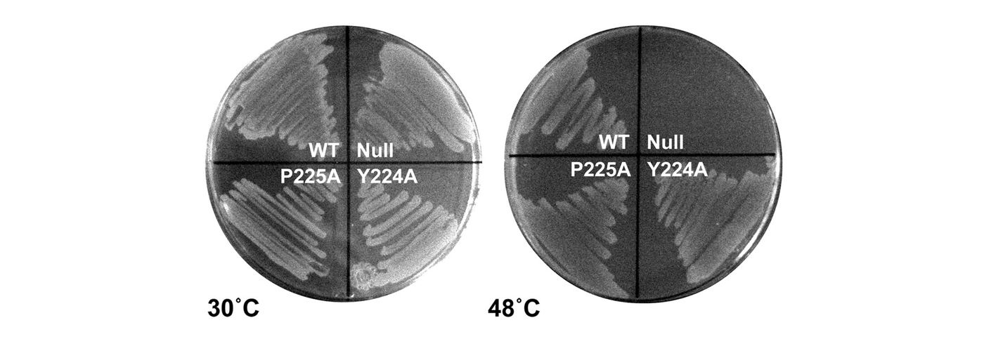
Figure 5.3: B. subtilis strains JH642 (wild-type; WT), RSA8 (Null), Y224A (divIB::cat::ermC amyE::divIB(Y224A)-cat; Y224A), and P225A (divIB::cat::ermC amyE::divIB(P225A)-cat; P225A) were streaked on LB plates supplemented with chloramphenicol at permissive (30˚C) and nonpermissive (48˚C) temperatures for the null strain.
here that result in poor insertion of DivIB into the cytoplasmic membrane without completely disrupting insertion may go unnoticed in this experimental system.
5.2.4 The domain of DivIB is crucial for function
In contrast to the point mutations made in the domain, a single point mutation in the region, Y246A, was sufficient to have a noticeable effect on colony formation ability at 48º C. Figure 5.4A illustrates that the Y246A strain (sector between 6 o’clock and 8 o’clock) at 30º C and 42º C (left and middle plate) is able to form colonies, however at 48º C (right plate), the streak of colonies appear to lyse (clear) after 24 hours of growth (it should be noted that even wildtype B. subtilis will lyse if left on an LB plate for 3-4 days). This is in contrast to the divIB strain, RSA8 (labeled NULL) which fails to grow at all at 48º C and wildtype (labeled WT) and fully complemented strains (labeled divIB) which do not lyse after only 24 hours of growth at 48º C.
The most likely cause of early lysis is filamentation due to a division defect in the Y246A strain. Light microscopy of strains RSA9 (labeled divIB), RSA29 (labeled Y246A) and RSA8 (labeled Null) grown for 3 hours at 48º C in liquid LB media are shown in figure 5.4B. There is no apparent filamentation of the Y246A strain compared to the wildtype divIB complemented strain RSA9. On the other hand, the divIB strain (RSA8) appears quite filamentous. While in liquid culture there does not appear to be a visible division defect that results from the Y246A mutation, this may not hold true for growth on solid media. Microscopic examination of cells after 24 hours of growth on plates could settle the matter. Curiously, however, it appears that the domain is significantly more
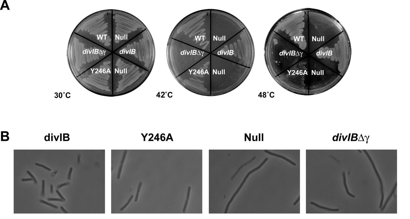
Figure 5.4: (A) B. subtilis strains JH642 (wild-type; WT), RSA8 (Null), RSA9 (divIB::cat::ermC amyE::divIB-cat; divIB), Y246A (divIB::cat::ermC amyE::divIB(Y246A)-cat; Y246A) and RSA29 (divIB::cat::ermC amyE::divIB-cat; divIB) were streaked on LB plates supplemented with chloramphenicol at permissive (30˚C) and the nonpermissive (42º C and 48˚C) temperatures for the null strain. (B) Micrographs of the strains in (A) grown in liquid culture for 3 hours.
sensitive to mutagenesis than the domain. This may, in part, explain why all known point mutations in the DivIB/FtsQ family map to this extreme C-terminal end.
Complementation of RSA8 with a divIB gene with a complete deletion of the domain of DivIB (RSA29, divIB) forms colonies at 30º C, fails to form colonies at 48º C, while early lysis appears to occur at 42º C (figure 5.4A, sector 8 o’clock to 10 o’clock). Microscopic examination of this strain after three hours of growth at 48º C in liquid culture reveals a filamentous phenotype (figure 5.4B, panel labeled divIB) and appears similar to the divIB null strain RSA8 (figure 5.4B, panel labeled Null). These results indicate that deletion of the entire domain has a severe effect on division. This combined with the single point mutation at Y246A furthermore indicates that the domain is surprisingly far more crucial for division than the domain. At least, the functional role of the domain cannot be compensated by high levels of DivIB protein as suggested may be the case for the domain in section 5.2.3.
5.2.5 Suppressor mutations of domain mutants?
In figure 5.4A, (48º C plate, sector divIB) several spots of colony formation can be seen, particularly towards the edge of the plate. These colonies were routinely observed. In addition, colonies were also observed when growing colonies of RSA8 (divIB) at 48º C, although less frequently (data not shown). While at present these colonies have not been verified as true spontaneous suppressor mutations of divIB mutations, this is a likely explanation for the presence of these colonies. If this were true, the fact that a divIB strain more readily generates suppressor mutants than a divIB strain indicates an important role of the DivIB protein outside of the region; a role that is harder to suppress in its absence than simply the absence of the domain alone. Suppressor mutations are likely to arise in genes for proteins that directly interact with the initial mutated protein. This suggests that the domain has a direct interaction with a protein that is readily mutated and results in a suppression of the divIB phenotype. In turn, the fact that suppressor mutations can arise, although less frequently in a divIB strain, indicates that some part of the DivIB protein outside of the domain most likely interacts with other proteins as well.
5.3 Discussion and Conclusions
The results presented here suggest the conserved surface of DivIB is either malleable or not critical for protein-protein interacts, at least under the conditions tested above. This is difficult to reconcile with the fact that over a wide range of species these surfaces do appear to be evolutionarily conserved. Why would such an area remain under selective pressure? While the inability to generate a phenotype from single point mutations in this region may be explained by the relative abundance of the DivIB protein in B. subtilis, this abundance should also lead to a drop in the selective pressure to maintain these surfaces. Most likely, the selective pressure to maintain these surfaces was exerted by forces of nature that were not replicated by the plating assay used above. The development of more sensitive assays for disruption of DivIB function may be more enlightening in the future.
While these surfaces may be critical for a function that is yet to be established, the failure to easily disrupt division by lesions of this surface suggests it may not be a good target for antibiotic development. This is particularly the case for bacterial species like B. subtilis, in which the DivIB protein is highly abundant. Alternatively, FtsQ, in E. coli, is a relatively scarce protein with approximately 22 copies per cell (Carson, Barondess et al. 1991); thus, targeting of FtsQ with rational drug design may be more productive.
Previous studies (reviewed in Section 1.2.3.3) have suggested that the C-terminal domain is sensitive to mutagenesis. Furthermore, this thesis has again highlighted the domain of the DivIB/FtsQ family of proteins as critical for function. How can such a relatively small region with apparently no structure in the absence of other divisomal proteins, in contrast to the larger and more rigidly structured domain, have such a vital role to play in cell division? Furthermore, if the role of DivIB is to stabilize the unstable FtsL and DivIC proteins, how is it possible that the domain of DivIB performs this function? It is clear that the domain is an attractive target for antibiotic development. Progress in this endeavor can only be made through: (i) finding binding partners for the domain and (ii) structural determination of the complex.