3. Interactions of DivIB, FtsL and DivIC
3.1 Introduction
The primary goal of this study was to structurally characterize DivIB, FtsL, and DivIC and to investigate their possible interactions with one another. First, concentrated and pure forms of DivIB, FtsL and DivIC were needed for biochemical and biophysical studies. We also decided it would be of great benefit to work with homologous proteins from two different Bacillus species, namely B. subtilis and G. stearothermophilus (a mild thermophile). B. subtilis was chosen because, in relation to cell division, it is the best studied Gram positive bacterium. G. stearothermophilus is a close relative of B. subtilis but it is a thermophile, and thermophilic proteins have often proved better than their mesophilic counterparts for biophysical studies. Because G. stearothermophilus is a close relative of B. subtilis we envisioned that structural studies of G. stearothermophilus proteins could be used to guide in vivo studies in B. subtilis which, unlike G. stearothermophilus, is amenable to genetic manipulation.
While the genes for DivIB, FtsL and DivIC have been previously described in B. subtilis and identified in other Bacillus genomes, none of these three proteins had been identified in the G. stearothermophilus genome prior to this thesis. In order to find these sequences, a TBLASTN search was conducted on the unfinished G. stearothermophilus genome (see: http://www.genome.ou.edu/bstearo.html) using the B. subtilis amino acid sequence for each protein as a query sequence. In the case of FtsL and DivIC a single contig was returned containing an entire open reading frame of the G. stearothermophilus ortholog. For DivIB, two contigs were returned that corresponded to the N-terminal region and the C-terminal region of B. subtilis DivIB with no sequences matching the central coding region. Fortunately, this did not prevent the design of primers to match the 5’ and 3’ ends of the coding sequence, which allowed PCR amplification of the entire DivIB gene from G. stearothermophilus.
3.2 Results
3.2.1 Sequence alignments
Figure 3.1 shows sequence alignments generated by the ClustalW program {Chenna, 2003 #177} for FtsL (upper panel) and DivIC (lower panel). Figure 3.2 shows the ClustalW alignment for DivIB. There is very little sequence conservation across species, however the level of conservation between Bacillus species and especially between B. subtilis and G. stearothermophilus, is much higher. Specifically, FtsL from B. subtilis and G. stearothermophilus are 38% identical, while the percentage homology increases to 59% when conservative substitutions are considered. For DivIC, the level of identity is 37% and the level of conservation is 58%. DivIB from B. subtilis is 38% identical with its G. stearothermophilus ortholog, and the level of conservation is 56%.
The transmembrane segments were identified as a sequence of 18-21 hydrophobic amino acids and are marked by solid bars in Figure 3.1. Previous biochemical work (Harry, Stewart et al. 1993) has established that the N-terminal portion of all three proteins resides in the cytoplasm hence the majority of the protein for all three proteins from all species resides in the extracytoplasmic space. All known deleterious point mutations for all three proteins lie in the extracytoplasmic regions (see Chapter 1), suggesting that these
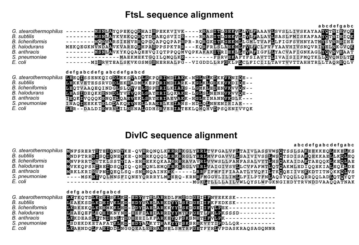
Figure 3.1: Alignment of FtsL (A) and DivIC (B) from G. stearothermophilus, B. subtilis, B. licheniformis, B. halodurans, B. anthracis, Streptococcus pneumoniae (all Gram positive eubacteria), and E. coli (a Gram negative eubacterium). The heptad repeats of the coiled-coil regions are marked with the customary “abcdefg” nomenclature above each alignment. The transmembrane regions are marked with a solid bar below each alignment.
domains represent the functionally important regions of these proteins. The extracytoplasmic regions for all three proteins from both B. subtilis and G. stearothermophilus were clearly identified based on these alignments, and these regions were subcloned for overproduction of the recombinant protein domains (see Section 3.2.2).
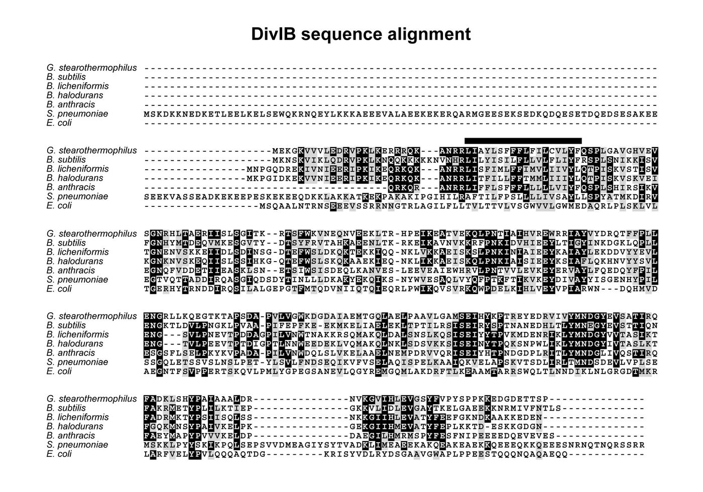
Figure 3.2: Alignment of DivIB from G. stearothermophilus, B. subtilis, B. licheniformis, B. halodurans, B. anthracis, Streptococcus pneumoniae (all Gram positive eubacteria), and E. coli (a Gram negative eubacterium). The transmembrane regions are marked with a solid bar above the alignment.
As discussed in Sections 1.2.3.4 and 1.2.3.5, both FtsL and DivIC contain putative coiled-coil domains. These have been indicated in Figure 3.1 by a heptad repeat of “abcdefg” where the ‘a’ and ‘d’ positions are typically occupied by hydrophobic residues {Junius, 1996 #178} that form the interhelical interface of the coiled coil. While the coiled-coils present in FtsL and DivIC from B. subtilis are apparently non-ideal (several ‘d’ positions are not occupied by leucine or other hydrophobic residues) the deviant residue pattern is preserved across all Bacillus species. This is suggestive of a conserved, but atypical, coiled coil structural motif across Bacillus species. The purpose of such an unusual coiled coil remains unknown.
3.2.2 Subcloning of the extracytoplasmic regions of FtsL, DivIC and DivIB.
The extracytoplasmic domains of all three proteins were directly cloned from chromosomal DNA from B. subtilis (strain 168) and G. stearothermophilus (strain NCB blah blah). The start point for each extracytoplasmic domain was easily identified based on the transmembrane regions shown in Figures 3.1 and 3.2. Using the designed forward and reverse primers from Table 2.1 with the appropriate chromosomal DNA resulted in a major dsDNA product of the appropriate size. Contaminating bands were usually present; however, these did not interfere with the cloning as they were significantly different in size and represented a minority of staining in the gel (i.e., they were minor products ofht epCR reaction). Bands of the correct size were excised from the gel, digested with BamHI and EcoRI restriction enzymes, then purified and ligated into pre-cut pGEX-2T vector. Ligation into pGEX-2T in this way results in a translational fusion between the gene for Schistosoma japonicum glutathione S-transferase (GST) protein and the coding sequence of the PCR product with an intervening thrombin cleavage site. Transformation of E. coli DH5 with the ligated vectors typically resulted in excess of 50 colonies on LB plates supplemented with ampicillin. A negative control of BamHI/EcoRI-cut pGEX-2T vector without ligation usually resulted in 0-2 colonies forming, indicating that this cloning strategy was highly successful.
Transformants were directly screened for their ability to overproduce fusion proteins of the correct size when induced with IPTG. Four single colonies from each transformation were grown in LB media and induced at mid-log phase with IPTG for 2 hours; samples were taken before and after induction with IPTG. These samples were run on SDS-PAGE and stained with coomassie blue. Successful transformants were identified by overproduction of protein of the appropriate molecular mass. Typically, 3-4 colonies overproduced protein with the correct molecular mass. A single successful transformant was randomly selected and plasmids prepared for DNA sequencing analysis. Once DNA sequencing had confirmed the correct DNA sequence was present the vector received a name (pSAR##; where ## corresponds to an index number) and was used to transform E. coli strains DH5 and BL21. The list of protein constructs, along with their matching plasmid names and strain numbers is given in Table 3.1.
Table 3.1 List of protein constructs, their corresponding plasmid names, strain numbers, and the molecular mass of the recombinant protein after thrombin cleavage of the GST fusion protein. * Note that each of these recombinant proteins contains a non-natural Gly-Ser at the N-terminus which is a vestige of the thrombin cleavage site.
| ProteinProteinProtein | Molecular Mass* Molecular Mass* Molecular Mass* (Da)(Da)(Da) | Plasmid Name Plasmid NamePlasmid Name (pGEX-2T derivative)(pGEX-2T derivative)(pGEX-2T derivative) | DH5 Strain #DH5DH5 Strain # Strain # | BL21 Strain #BL21 Strain #BL21 Strain # |
|---|---|---|---|---|
| Bsub-FtsL54–117Bsub-FtsLBsub-FtsL54–11754–117 | 7318.37318.37318.3 | pSAR11pSAR11pSAR11 | 209209209 | 210210210 |
| Bsub-DivIC58–125Bsub-DivICBsub-DivIC58–12558–125 | 7936.97936.97936.9 | pSAR12pSAR12pSAR12 | 211211211 | 212212212 |
| BSub-DivIB54-263BSub-DivIBBSub-DivIB54-26354-263 | 23929.123929.123929.1 | pSAR20pSAR20pSAR20 | 317317317 | 318318318 |
| Bste-FtsL63–126Bste-FtsLBste-FtsL63–12663–126 | 7527.47527.47527.4 | pSAR13pSAR13pSAR13 | 213213213 | 214214214 |
| Bste-DivIC58–123Bste-DivICBste-DivIC58–12358–123 | 7977.77977.77977.7 | pSAR14pSAR14pSAR14 | 215215215 | 216216216 |
| Bste-DivIB46-261Bste-DivIBBste-DivIB46-26146-261 | 24126.324126.324126.3 | pSAR15pSAR15pSAR15 | 233233233 | 234234234 |
3.2.3 Purification of extracytoplasmic domains of FtsL, DivIC and DivIB.
Each fusion protein was first purified from cell lysates using glutathione affinity chromatography as outlined in section 2.x, then the protein of interest was liberated by on-column thrombin cleavage and purified further using ion exchange chromatography. The ion exchange chromatograms — especially for FtsL and DivIC — were quite complicated and comprised many peaks that are presumably fragments of full length protein. Indeed, the purification gels show that both B. subtilis and G. stearothermophilus FtsL and DivIC are considerably heterogeneous when eluted from the glutathione affinity matrix (Figure 3.3, lanes 6). However, there was always a dominant peak in the FPLC
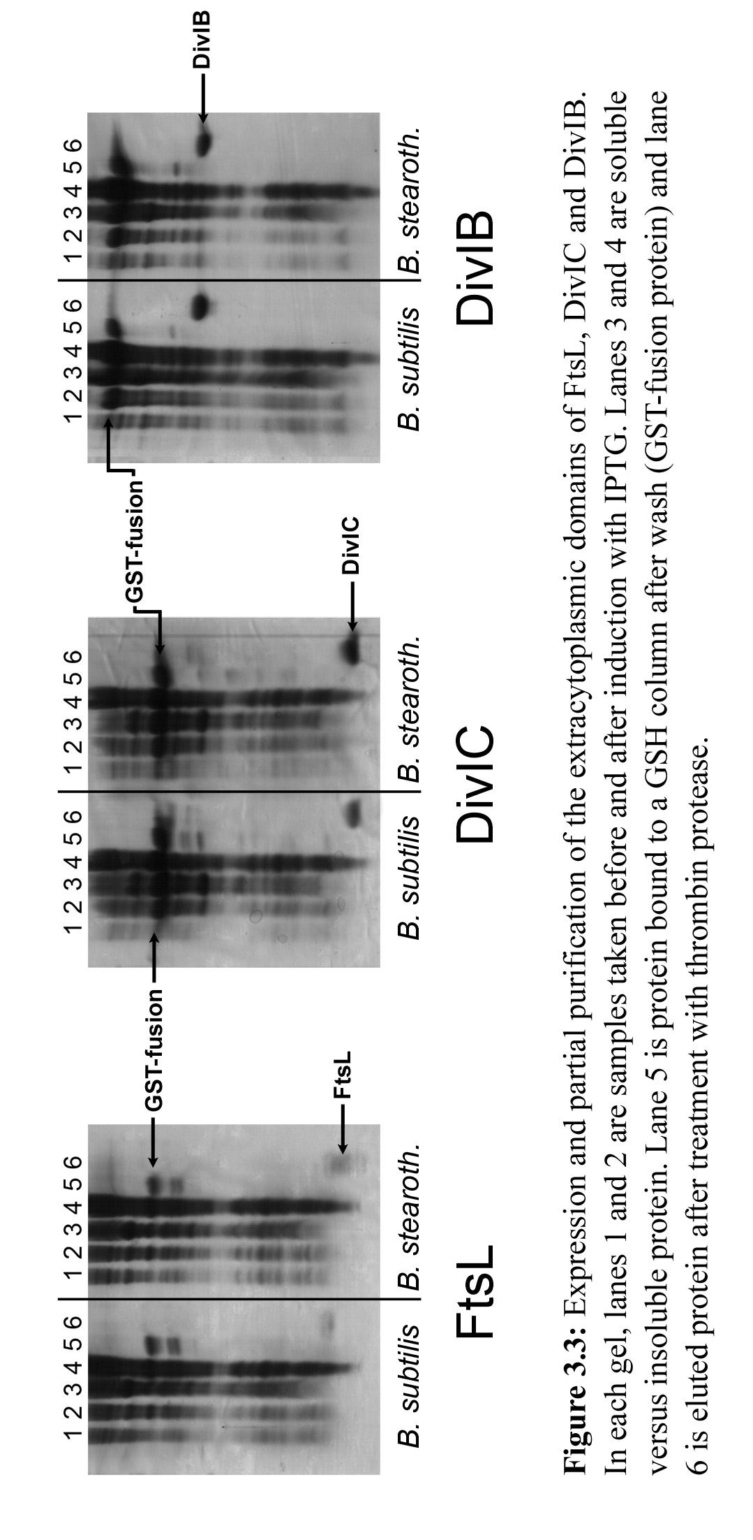
chromatograms which when collected and subjected to SDS-PAGE appeared to have a mass corresponding to the full-length construct.
The identity of the purified recombinant proteins was confirmed using mass spectrometry. For MALDI mass spectral analysis, ion-exchange-purified proteins were subjected to reverse phase HPLC to remove salts which interfere with the MALDI procedure. When these proteins where rerun on reverse phase HPLC, they yielded a single peak, confirming purity. MALDI mass spectral analysis verified that each construct was of the correct molecular weight to within 1-2 Da. It should be noted that, despite the problems with overproduction of what appear to be quite unstable proteins, pure forms of each protein were obtained using methods that are expected to maintain the native fold of the protein. Apart from molecular mass determination by MALDI, all experiments were performed with proteins purified in the native state.
3.3 Biophysical Analysis of FtsL, DivIC and FtsL/DivIC interactions
The experiments reported here used protein constructs corresponding to the entire predicted extracytoplasmic domains of B. subtilis FtsL (residues 54–117; Bsub-FtsL54-117), B. subtilis DivIC (Bsub-DivIC58–125), G. stearothermophilus FtsL (Bste-FtsL63–126), and G. stearothermophilus DivIC (Bste-DivIC58–123); the cloning and purification of the domains is described in Sections 3.2.2 and 3.2.3. Even though these domains were overproduced in a protease-deficient strain of E. coli (BL21, lon–, ompT–) and protease inhibitors were used during the purification procedure, there was noticeable degradation of all proteins (except G. stearothermophilus DivIC) after storage for 1 week at 4º C. Thus, all subsequent experiments were conducted within 1–2 days of protein purification, and SDS–PAGE gels recorded after the experiments revealed no significant protein degradation.
3.3.1 CD analysis of purified extracytoplasmic FtsL and DivIC
Far-UV circular dichroic (CD) spectra of each of these domains are shown in Figure 3.4. It is immediately apparent that the B. subtilis protein domains as well as Bste-FtsL63–126 are predominantly unstructured, containing large amounts of random coil (which yields a deep minimum at 199 nm) and very small amounts of -helical secondary structure (minima at 208 and 222 nm) (Woody 1996). The percentages of -helical secondary structure calculated from the ellipticities at 222 nm (Pelton and McLean 2000) were ~6% (Bsub-FtsL54–117), ~8% (Bsub-DivIC58–125) and ~12% (Bste-FtsL63–126). In marked contrast, the CD spectrum of Bste-DivIC58–123 is characteristic of a protein with significant -helical secondary structure; the percentage -helix calculated from the prominent minimum at 222 nm is ~40%. Interestingly, ~40% of the primary structure of Bste-DivIC58–123 is predicted to be a coiled-coil; however, multi-angle laser light scattering (MALLS) experiments on this construct have shown it exists as a monomer. Indeed, all four proteins are monomeric in solution as judged by MALLS (see Table 3.2). Although each molecular mass prediction is slightly higher than the true molecular mass (between 7.6% and 11% higher) this error is most likely the result of a lack of sensitivity in the scattering measurements as these proteins are relatively small and they scatter light
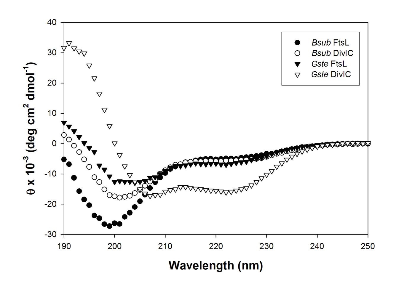
Figure 3.4: Far-UV circular dichroic spectra of the extracytoplasmic domains of B. subtilis and G. stearothermophilus FtsL and DivIC. The abscissa indicates mean residue ellipticity (). Note that only G. stearothermophilus DivIC lacks a dominant random coil signature (minimum at 199 nm).
poorly. Alternatively, the slightly higher than monomeric measurements may reflect a small tendency to non-specifically aggregate under the chosen buffer conditions.
The high percentage of random coil structure is reflected in the thermal denaturation profiles of these protein domains (see Figure 3.5). The thermal denaturation curve for Bsub-FtsL54–117 has a negative slope, which is characteristic of an almost completely random coil peptide (Woody 1992); therefore, we concluded that this protein domain is essentially completely unfolded even at 0º C. The thermal denaturation profiles for both
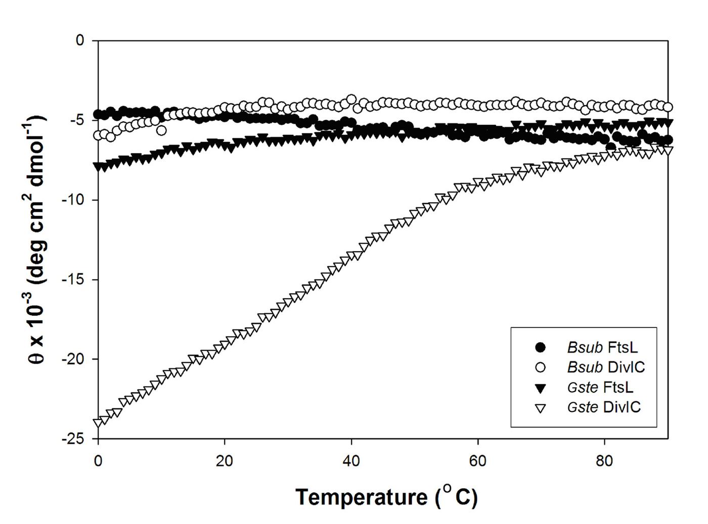
Figure 3.5: Thermal denaturation profiles of the extracytoplasmic domains of B. subtilis and G. stearothermophilus FtsL and DivIC. The abscissa indicates mean residue ellipticity (). Each domain is predominantly unfolded at 0º C with the exception of G. stearothermophilus DivIC.
Bsub-DivIC58–125 and Bste-FtsL63–126 reveal that they are completely unfolded by the time the temperature reaches 30º C, which indicates that both protein domains will be completely unfolded in vitro at the optimal growth temperatures for these two organisms. As anticipated from its higher -helical content, the thermal denaturation profile of Bste-DivIC58–123 reveals that it is more thermodynamically stable than the other three protein domains. However, the melting temperature (Tm, defined as the temperature at which the domains are half unfolded) is still <40º C, and the protein is almost completely unfolded at 60º C, the optimal growth temperature for G. stearothermophilus. It seems unlikely that the instability of these proteins results from the absence of their associated cytoplasmic and transmembrane domains as, at least in the case of B. subtilis DivIC, it has been shown that these regions can be replaced with the cytoplasmic and transmembrane domains of heterologous membrane proteins without loss of function (Katis and Wake 1999). Thus, it is concluded that the extracytoplasmic domains of DivIC and FtsL are both thermodynamically highly unstable and are likely to be rapidly degraded in vivo unless stabilized by interaction with other divisomal proteins.
Table 3.2: Comparison of the molecular mass of FtsL and DivIC predicted on the basis of their primary structure with the molecular mass determined using MALLS. Each construct appears to be monomeric in solution. * Note that each of these recombinant proteins contains a non-natural Gly-Ser at the N-terminus, which is a vestige of the thrombin cleavage site.
| ProteinProteinProtein | Predicted Molecular Mass* Predicted Molecular Mass* Predicted Molecular Mass* (Da)(Da)(Da) | Molecular Mass as determined by MALLS Molecular Mass as determined by MALLS Molecular Mass as determined by MALLS (Da)(Da)(Da) | Percentage errorPercentage errorPercentage error (%)(%)(%) |
|---|---|---|---|
| Bsub-FtsL54–117Bsub-FtsLBsub-FtsL54–11754–117 | 7318.37318.37318.3 | 8157 ± 598157 ± 598157 ± 59 | 111111 |
| Bsub-DivIC58–125Bsub-DivICBsub-DivIC58–12558–125 | 7936.97936.97936.9 | 8543 ± 888543 ± 888543 ± 88 | 7.67.67.6 |
| Bste-FtsL63–126Bste-FtsLBste-FtsL63–12663–126 | 7527.47527.47527.4 | 8226 ± 1088226 ± 1088226 ± 108 | 9.29.29.2 |
| Bste-DivIC58–123Bste-DivICBste-DivIC58–12358–123 | 7977.77977.77977.7 | 8734 ± 558734 ± 558734 ± 55 | 9.59.59.5 |
3.3.2 FtsL and DiviC Interaction studies
In Section 3.3.1 it was established that Bsub-FtsL54–117, Bsub-DivIC58–125 and Bste-FtsL63–126 are essentially unfolded proteins which do not self-associate, since specific homo-interactions of these proteins would be accompanied by regular secondary structure (which was not detected using CD) and the MALLS data indicates that the proteins were predominantly monomeric. Bste-DivIC58–123 is clearly monomeric as well as judged by MALLS data. Thus, it appears none of these proteins is able to self-associate via their predicted coiled-coil regions or any other part of the protein. One of the fundamental questions posed by the genetic data described in the introduction to this thesis is — do FtsL and DivIC hetero-interact with each other via their predicted coiled-coil regions? A report in the literature suggested that the extracytoplasmic domains were sufficient to support such an interaction (Sievers and Errington 2000). However, it was decided to reinvestigate this hypothesis using a range of biochemical and biophysical techniques.
3.3.2.1 Circular Dichroic Analysis of Putative FtsL-DivIC interaction.
As the CD spectra of Bsub-FtsL54–117 and Bsub-DivIC58–125 indicated that both proteins are predominantly random coil, it was reasoned that any interaction between these proteins should be accompanied by an increase in structure, which would be reflected in the CD spectrum of the complex. An example of such a structural transition upon complex formation is the dramatic random coil to -helix transition that occurs when the DNA-binding domains of the transcriptional activators c-Jun, c-Fos and GCN4 bind their cognate enhancer elements (Patel, Abate et al. 1990; Weiss, Ellenberger et al. 1990). Figure 3.6A shows CD spectra of 10 M solutions of Bsub- FtsL54–117 (blue) and Bsub-DivIC58–125 (red) and an equimolar mixture of the two protein domains (both proteins at 10 M, slate). It can be seen that the CD spectrum of the mixture is not significantly different from the mathematical sum of the spectra of the individual proteins (green). In other words, the structure of neither protein is affected by the presence of the other; both proteins remain predominantly unstructured. Thus, the CD experiments argue strongly against an interaction between these two protein domains, as it is difficult to envisage a complex in which both proteins remain predominantly unstructured. Furthermore, it seems very unlikely that the proteins would interact without experiencing any structural perturbations relative to their uncomplexed state. A similar series of experiments was performed using the G. stearothermophilus protein domains (Figure 3.6B). Once again,
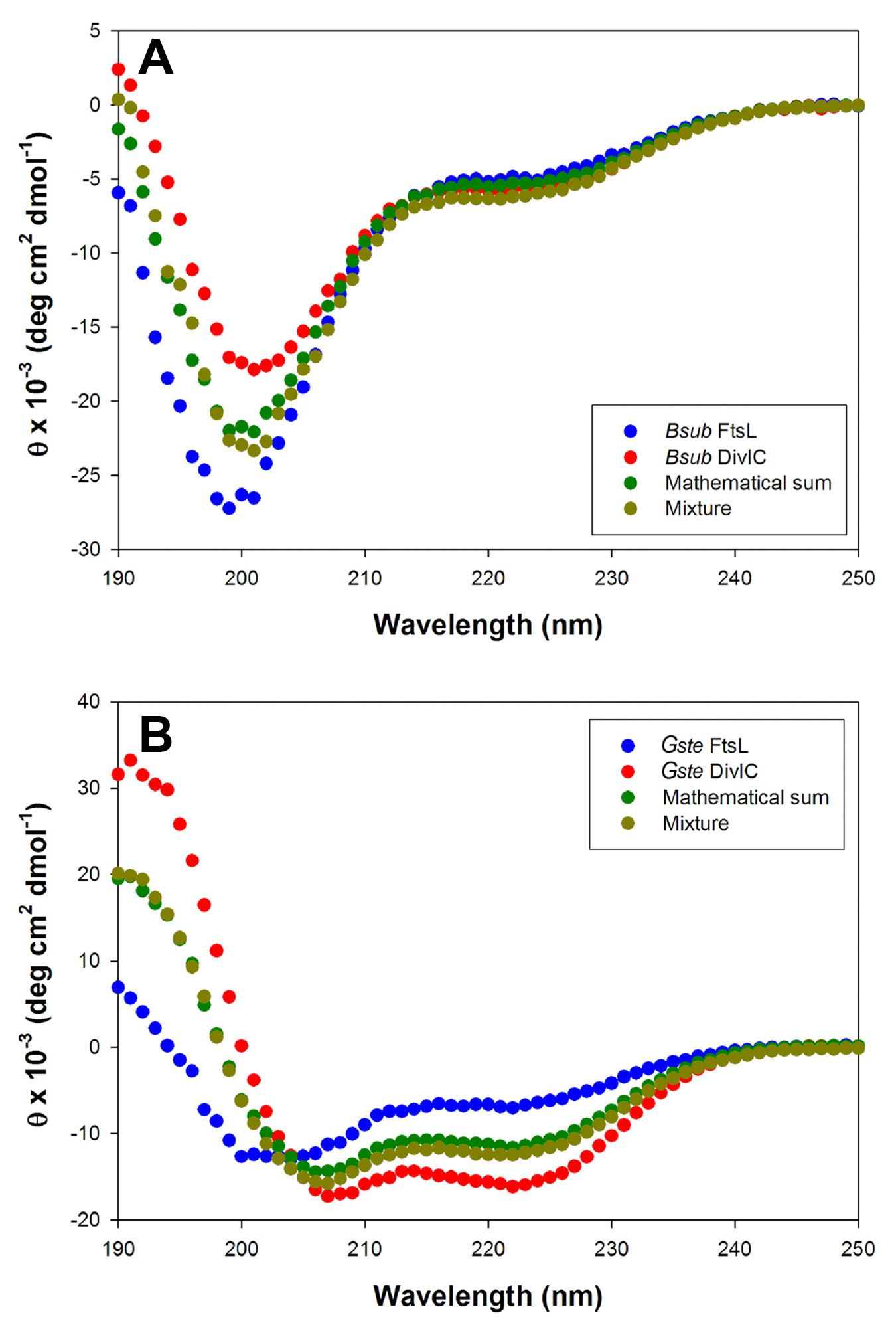
Figure 3.6: (A). Far-UV CD spectra of the extracytoplasmic domains of B. subtilis (Bsub) FtsL (blue) and DivIC (red), as well as an equimolar mixture of the two proteins (orange). The mathematical sum of the spectra of the individual domains (green) is almost identical to the spectrum of the equimolar mixture of the two proteins (slate). (B). Far-UV CD spectra of the extracytoplasmic domains of G. stearothermophilus (Gste) FtsL (blue) and DivIC (red), as well as an equimolar mixture of the two proteins (slate). The mathematical sum of the spectra of the individual domains (green) is almost identical to the spectrum of the equimolar mixture of the two proteins (slate).
the CD spectrum of a 10 M mixture of the two proteins (slate) was not significantly different from the mathematical sum of the spectra of the individual proteins (green), indicating that the structure of neither protein domain is perturbed by the presence of the other. Thus, the CD data argue strongly against a direct interaction between DivIC and FtsL.
3.3.2.2 Ion exchange analysis of putative FtsL-DivIC interation.
The predicted isoelectric points of Bsub-FtsL54–117 (pI ~8.9) and Bsub-DivIC58–125 (pI ~5.5) are markedly different, which enables them to be readily separated using ion exchange chromatography. Under the conditions used to obtain the chromatograms shown in Figure 3.7 (pH 6.7), Bsub-FtsL54–117 was significantly retained on a MonoS cation exchange column (Figure 3.7A, centre), whereas Bsub-DivIC58–125 eluted in the void (Figure 3.7A, bottom). It was reasoned that, if there was an interaction between these two protein domains, then the resultant complex would have an intermediate pI (e.g. the predicted pI of a 1:1 complex is 8.0) and therefore should elute with a retention time between that of the individual protein domains. However, application of an equimolar mixture of the two protein domains to the MonoS column yielded a chromatogram (Figure 3.7A, top) that only contained peaks at the positions expected for uncomplexed Bsub-FtsL54–117 and Bsub–DivIC58–125, which argues against an interaction between the two proteins.
It is conceivable, however, that a binary complex with a stoichiometry highly biased towards one of the two domains would elute close to the retention time of that domain. Fortunately, despite their very similar molecular weights, Bsub-FtsL54–117 and Bsub-DivIC58–125 migrate differently during SDS–PAGE (Figure 3.7B, lanes 6 and 7) and, to examine the protein content of each peak from the chromatogram. It is clear from the gel shown in Figure 3.7B that the void peak (VP) in the chromatogram of the mixture (see lane 4) contains only DivIC, whereas the retained peak (RP; see lane 5) only contains FtsL. Thus, it can be safely concluded that, under these conditions, there is no interaction between the extracytoplasmic domains of B. subtilis FtsL and DivIC.
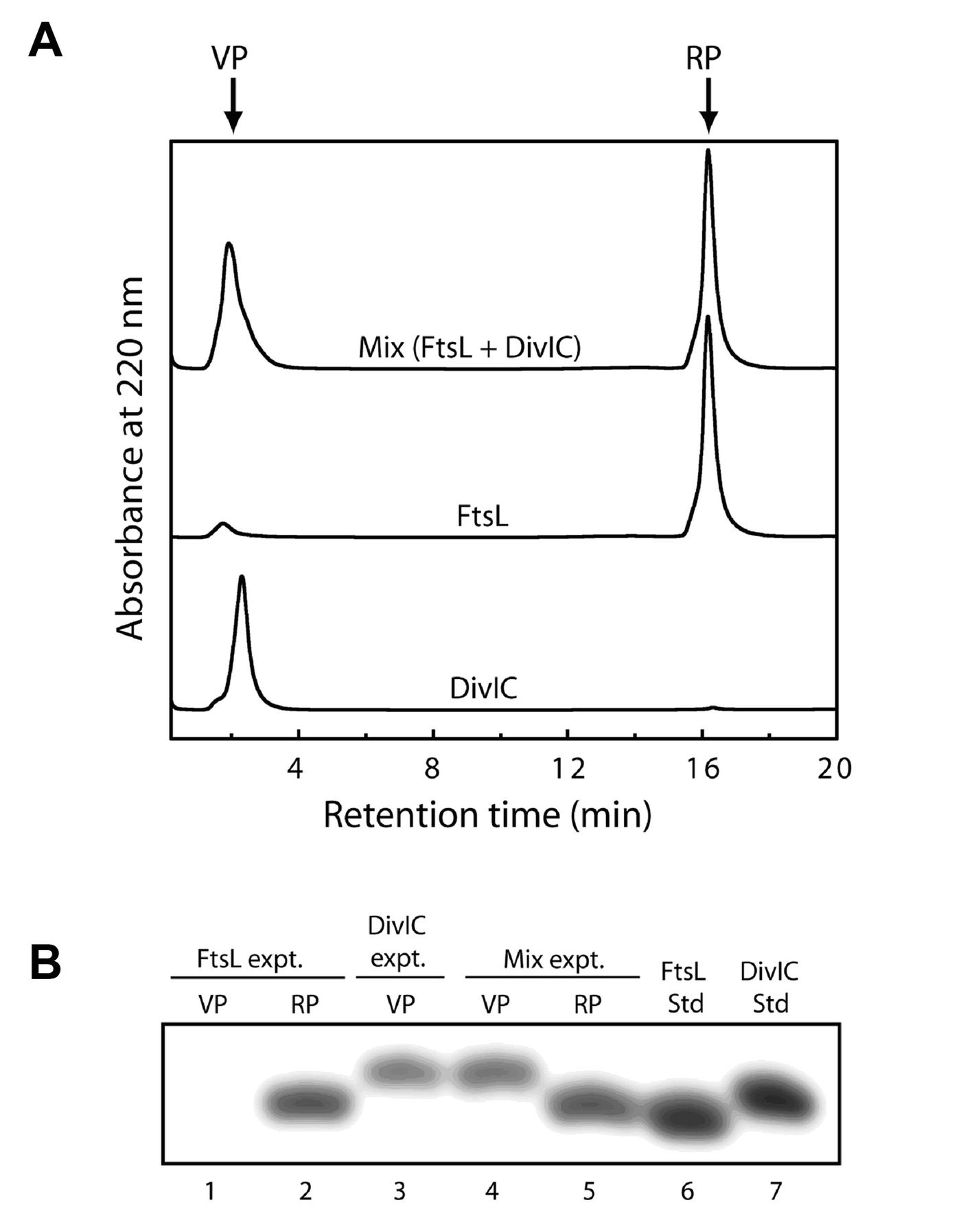
Figure 3.7: (A) Chromatograms obtained from applying the extracytoplasmic domains of B. subtilis DivIC (bottom), FtsL (middle), and an equimolar mixture of the two domains (top) to a Pharmacia MonoS cation exchange column. FtsL is significantly retained under these experimental conditions, whereas DivIC elutes close to the column void. Note that no additional peaks are apparent in the mixture of the two domains. (B) SDS–PAGE gel analysis of the retained peak (RP) and void peak (VP) from each of the chromatograms shown in (A). Note that the void peak in the equimolar mixture contains only DivIC and the retained peak contains only FtsL.
3.3.2.3 NMR analysis of the putative FtsL-DivIC interaction.
The 15N-edited heteronuclear single-quantum coherence (HSQC) spectrum of a uniformly 15N-labeled protein contains a single crosspeak for each backbone amide group as well as pairs of crosspeaks for each Gln and Asn sidechain amide moiety. The 1H and 15N resonance frequencies of these cross-peaks are exquisitely sensitive to the chemical environment of the amide group and can be substantially altered by the proximal binding of even small ligands (Maciejewski, Shin et al. 2001). Thus, it was reasoned that the HSQC spectrum of 15N-labeled Bsub-FtsL54–117 should be markedly altered if it forms a complex with Bsub-DivIC58–125, and vice versa. Figure 3.8A and 3.8B shows the 15N-edited HSQC spectra of uniformly 15N-labeled Bsub-FtsL54–117 and Bsub-DivIC58–125, respectively, each at a concentration of 230 M. As anticipated from the CD experiments reported above, most of the amide–proton chemical shifts are concentrated in the expected range (8.0–8.5 p.p.m.) for largely random coil proteins (Wuthrich 1986). Figure 3.8C shows the HSQC spectrum acquired from an equimolar mixture of the labelled protein domains (each domain at 230 M). Figure 3.8D shows an overlay of the HSQC spectra of the individual proteins on the spectrum of the equimolar mixture. It can be seen that the spectrum of the protein mixture is essentially identical to the sum of the spectra of the individual protein domains; there is no difference in the resonance frequencies of any of the crosspeaks in either of the proteins when they are mixed compared with when they are alone. Given the extreme sensitivity of these resonance frequencies to even minor changes in the chemical environment, these data provide very strong evidence that there is not even a transient interaction between the extracytoplasmic domains of B. subtilis FtsL and DivIC.
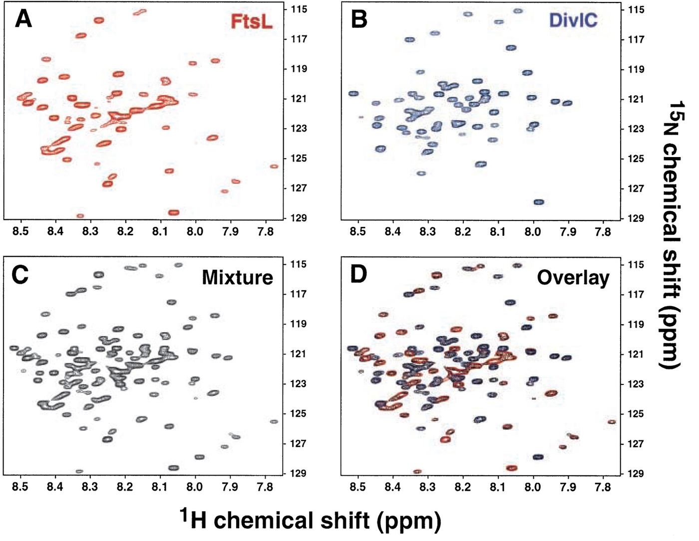
Figure 3.8: FtsL and DivIC do not interact directly. Two-dimensional HSQC NMR spectra of 15N-labeled extracytoplasmic domains of B. subtilis FtsL (A), DivIC (B) and an equimolar mixture of the two proteins (C). (D) The spectra of the two individual domains have been overlaid on the spectrum of the equimolar mixture, with preservation of colors. The near-perfect superposition of the individual spectra on the spectrum of the mixture indicates that the NMR spectrum of neither protein is perturbed by the presence of the other.
3.3.2.4 CD analysis in the presence of Lipids
Given the proximity of each of these protein domains to the cytoplasmic membrane it is entirely possible that interactions between the extracytoplasmic domains of FtsL and DivIC are mediated or stabilized by interactions with a lipid bilayer. Spontaneous formation of stable -helix in the presence of lipids, even for short peptides, has been reported previously (Szeto, Rowland et al. 2003). Addition of 500 M small unilamellar vesicles (SUVs, see section 2.3.8 for preparation of B. subtilis-like SUVs) to 10 M Bsub-FtsL54–117 or 10 M Bsub-DivIC58–125 (50:1 ratio) did not induce any apparent secondary structure in the individual proteins when assayed using CD spectropolarimetry, indicating that neither protein is able to interact with the SUVs in isolation (data not shown). In order to test if SUVs are sufficient to induce an interaction between FtsL and DivIC, 500 M SUVs were incubated with 10 M Bsub-FtsL54–117 and 10 M Bsub-DivIC58–125, then the mixture was analyzed using CD spectropolarimetry. Figure 3.9 shows CD spectra of a solution of 10 M solutions of Bsub-FtsL54–117 plus Bsub-DivIC58–125 in the absence (blue) or presence (red) of 500 M SUVs. The two spectra align almost perfectly, indicating that SUVs do not induce conformational alterations in these proteins, which in turn implies that lipids are not sufficient to stabilize an interaction between FtsL and DivIC.
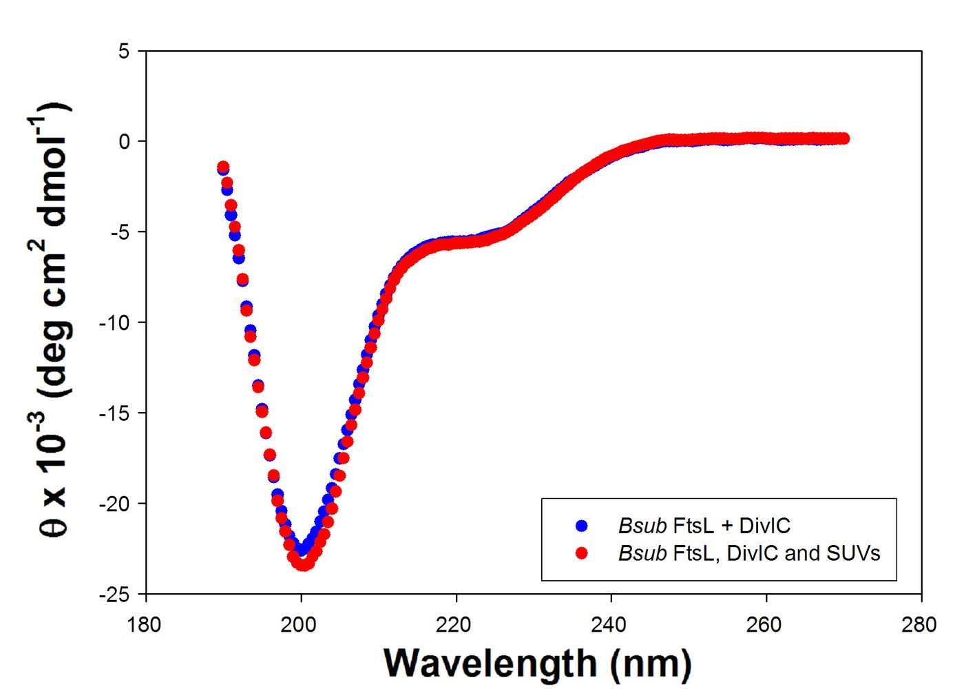
Figure 3.9: Far-UV CD spectra of the extracytoplasmic domains of B. subtilis (Bsub) FtsL and DivIC in the absence (blue) and presence (red) of B. subtilis-like SUVs. The presence of SUVs does not significantly alter the spectrum of FtsL and DivIC which indicates that lipids do not cause structural changes in either protein or promote their association.
3.3.2.5 FtsL and DivIC do not interact.
Despite a previous report that FtsL and DivIC interact directly and exclusively by their extracytoplasmic domains in a native gel experiment (Sievers and Errington 2000), this result was unable to be replicated here using several other techniques to probe for protein-protein interactions. During the initial phase of this work, an attempt was made to replicate the native gel experiment of Sievers and Errington. Despite using an identical protocol and protein constructs as Sievers and Errington, this result was unable to be replicated in this thesis. Indeed, initial native gel experiments were a failure as FtsL would not even migrate into the native gel at a running pH of 8.7 (the pH at which the original Sievers and Errington experiment was run). This is not surprising as the pI of FtsL is 8.9 – a physical property that would mean FtsL would never run into the gel. Upon adjusting the pH of the native gel to 9, FtsL was seen to run into the gel, however still no interaction with DivIC was observed. Given this result and the results presented here, it is concluded that the original observation, of Sievers and Errington, that FtsL and DivIC can directly interact with each other in the absence of other divisomal components was in error.
3.3.3 Protein-protein interactions between DivIB, FtsL and DivIC.
Given the dependency of FtsL stability on DivIB (Katis, Wake et al. 2000) it is entirely possible that the DivIB protein can either interact with FtsL or stabilize a complex of FtsL and DivIC. In order to test for this possibility MALLS analysis in tandem with gel filtration chromatography was employed for testing protein-protein interactions. MALLS determines the molecular mass of proteins and the total molecular mass of protein complexes after elution from a size exclusion chromatography column. As such, a 1:1:1 stiochiometric complex of DivIB, FtsL and DivIC would expect to give rise to molecular mass predictions of the sum total of each individual molecular mass (in the case of the B. subtilis proteins, that is a sum total molecular mass of 39184.3 gmol-1).
3.3.3.1 MALLS analysis of interacts between DivIB, FtsL and DivIC.
In order to test for potential interactions between DivIB, FtsL and DivIC via MALLS, it was necessary to establish the MALLS properties of each protein before mixing. Table 3.3 (column 3) summarizes the molecular mass determinations for each protein construct via MALLS when run individually. Apart from the results for DivIB, these results have already been discussed in section 3.3.1. I need to briefly discuss the results for DivIB here – one sentence should do it. Figure 3.10 (panel A – solid line) displays the size-exclusion chromatogram of a 1:1:1 mix of DivIB, FtsL and DivIC (40 M each). Molecular mass predictions (solid circles) have been generated for two observed peaks after chromatography. Panel B contains a repeat experiment, this time using the protein constructs from G. stearothermophilus (conducted at a 1:1:1 ratio of each protein at 60 M). Based on the molecular mass predictions for each protein peak it is apparent that the first peak in each trace (peak on the left side) represents most likely well resolved DivIB. The second peak, based also on molecular mass prediction and the apparent width of the peak appears to contain a mix of FtsL and DivIC protein. The molecule mass predictions for each peak appear in table 3.2. It is apparent from table 3.2 that the first peak in each experiment (DivIB) most likely contains DivIB protein alone and the second peak is FtsL unresolved from DivIC.
Amazingly, in the case of B. subtilis DivIB, the predicted molecular mass has fallen upon addition of FtsL and DivIC (-9.0%). This may be the result of poor resolution of DivIB from the second peak that contains smaller proteins and may bias the predictions to lower values. On the other hand, the molecular mass prediction for the second peak (FtsL and DivIC) is 8.5% higher when preincubated with DivIB – this is based on the assumption that FtsL and DivIC do not interact and the second peak would have predictions for the average of the molecular masses of FtsL and DivIC alone. Once again, the slight increase in molecular mass prediction here could be the result of poor resolution from the higher molecular mass DivIB peak. In the case of the G. stearothermophilus proteins (Figure 3.10B) there is essentially no difference between the first peak and DivIB when run alone (1.1% difference), suggesting this peak is simply DivIB. The second peak has molecular mass predictions some 20% higher than expected for an equimolar mix of FtsL and DivIC. While one interpretation of this result is a small amount of DivIB is shifted to this peak, indicating an interaction (N.B peak separation in panel B is poorer than in panel A) a more likely interpretation is simply poorer separation between DivIB and FtsL plus DivIC. Also, a complex of DivIB, FtsL and DivIC would more likely elute in front (to the left) of the DivIB peak. Closer examination of the molecular mass predictions in panel B show a distinct gradient in the predictions for the second peak as well, with predictions rising to the left – a likely result of poor resolution from the peak preceding it. Taken together, these two results suggest there is no stable interaction between DivIB, FtsL and DivIC.
Table 3.3: Comparison of molecular masses determined by MALLS for DivIB, FtsL and DivIC from B. subtilis and G. stearothermophilus before and after mixing.
| ProteinProteinProtein | True molecular massTrue molecular massTrue molecular mass (gmol-1)(gmol(gmol-1-1)) | Molecular mass - individualMolecular mass - individualMolecular mass - individual (gmol-1)(gmol(gmol-1-1)) | Molecular mass - mixedMolecular mass - mixedMolecular mass - mixed (gmol-1)(gmol(gmol-1-1)) | Apparent change in molecular massApparent change in molecular massApparent change in molecular mass (%)(%)(%) |
|---|---|---|---|---|
| Bsub FtsLBsubBsub FtsL FtsL | 7318.37318.37318.3 | 8157 ± 608157 ± 608157 ± 60 | 9061 ± 659061 ± 659061 ± 65 | 8.58.58.5 |
| Bsub DivICBsubBsub DivIC DivIC | 7936.97936.97936.9 | 8543 ± 888543 ± 888543 ± 88 | ||
| Bsub DivIBBsubBsub DivIB DivIB | 23929.123929.123929.1 | 29982 ± 22829982 ± 22829982 ± 228 | 27286 ± 6127286 ± 6127286 ± 61 | -9.0-9.0-9.0 |
| Gste FtsLGsteGste FtsL FtsL | 7527.47527.47527.4 | 8226 ± 1088226 ± 1088226 ± 108 | 10178 ± 18710178 ± 18710178 ± 187 | 202020 |
| Gste DivICGsteGste DivIC DivIC | 7977.77977.77977.7 | 8735 ± 558735 ± 558735 ± 55 | ||
| Gste DivIBGsteGste DivIB DivIB | 24126.324126.324126.3 | 25145 ± 13425145 ± 13425145 ± 134 | 25420 ± 13025420 ± 13025420 ± 130 | 1.11.11.1 |
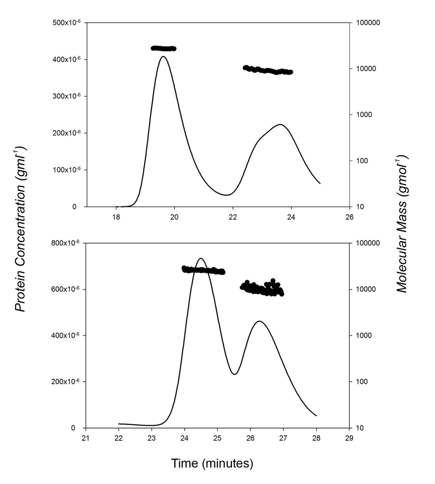
Figure 3.10: Size exclusion chromatography trace (solid line) of an equimolar mix of DivIB, FtsL and DivIC from B. subtilis (A) and G. stearothermophilus (B). Chromatography results in the separation of the proteins into two peaks. Each peak has the molecular mass estimated using multi-angle laser light scattering (MALLS) (solid dots). The peak to the left in each panel appears to be composed exclusively of monomeric DivIB protein while the peak to the right appears to be composed of a mix of monomeric FtsL and DivIC.
3.4 Discussion and Conclusions
During the course of this thesis, two papers where published in which stable in vitro interactions between DivIB/FtsQ, FtsL and DivIC/FtsB were reported; firstly, a paper describing this ternary interaction with E. coli proteins (Buddelmeijer and Beckwith 2004), and a second paper describing a ternary interaction with the proteins from Streptococcus pneumoniae (Noirclerc-Savoye, Le Gouellec et al. 2005). In each case, the experimental conditions are distinct from the conditions attempted here; the differences between these experiments give indications about what exactly is necessary and sufficient for DivIB, FtsL and DivIC to interact.
In the paper of Buddelmeijer and Beckwith (2004), an interaction between FtsL and FtsB (DivIC) was first indicated using a co-precipitated protocol in which an antibody for FtsL (-FtsL) was incubated with solubilized membrane protein (solubilized with 1% triton X-100). The antibody plus bound protein complexes were purified. -FtsL was found to co-precipitate FtsL (by western blot) and FtsB, indicating that FtsL and FtsB exist as a complex in solubilized membranes. The reverse was also established using an anti-FLAG tagged FtsB to co-precipitate FtsB-FLAG and FtsL. They also established that swapping the transmembrane domain of FtsL for that of an unrelated transmembrane protein disrupted the interaction, as did replacing the coiled-coil region of E. coli FtsL with that from H. influenzae FtsL. Swapping the transmembrane domain of FtsL for that of FtsQ resulted in poorer complex formation but did not obliterate it completely. Thus, the transmembrane and leucine-zipper motif are essential for this interaction. In addition, they also showed FtsQ (DivIB) was also part of this co-precipitated complex by western-blot, probing with anti-FtsQ antibodies.
While the results above suggest that FtsQ, FtsL and FtsB are able to form a complex, there is no indication that these proteins are sufficient to form a stable complex. It is entirely possible that other proteins – undetected by the specific nature of western-blot analysis – are present in the complex, perhaps performing as a vital component of the interaction between the remaining three proteins. The use of solubilized membranes and disruption of interactions by changes in the transmembrane region of FtsL suggest correct transmembrane sequences and/or membrane context are important for the interaction. In B. subtilis, it has been established that the transmembrane regions of DivIC and DivIB are not important for function (Katis and Wake 1999). If the nature of the interaction between these three proteins is constant between B. subtilis and E. coli, it becomes hard to reconcile the important of the FtsL transmembrane region in a ternary interaction with the fact the two other remaining transmembrane segments can be substituted for irrelevant (though still membrane-spanning-like) sequences. Indeed, in the case of E. coli it has been established that the specific amino-acid sequence of the transmembrane segment of FtsQ is not needed for function (Chen, Weiss et al. 1999). If the actual specific amino-acid sequence of the transmembrane region of FtsL in E. coli is not important for function, it may instead play a role in establishing or stabilizing a conformational state in the extracytoplasmic region – specifically, supporting -helical and coiled-coil structures in the region proximal to the membrane. Such a role for the transmembrane segment of FtsL could only manifest in the context of proper membrane insertion that may be mimicked in a detergent solubilized membrane preparation.
In the second paper (Noirclerc-Savoye, Le Gouellec et al. 2005), a ternary interaction was reconstituted in vitro in the absence of membranes, detergents and clearly demonstrated that the extracytoplasmic domains of DivIB, FtsL and DivIC are sufficient for stable interaction. A GST-fusion to DivIB was able to co-precipitate a mix of FtsL and DivIC forced to heterodimerize by addition of artificial coiled-coils at their N-terminal end (essentially where their transmembrane domains would normally be situated). Interestingly, the introduction of these artificial coiled-coils is essential for a ternary interaction to take place. In their absence (essentially a similar scenario to the results presented in section 3.3.3.1), no ternary interaction was observed; this in part verifies the result reported in section 3.3.3.1 of this thesis. While a stable interaction between DivIB, FtsL and DivIC is gratifying in terms of the genetic data discussed in the introduction, it remains an open question why forcing heterodimerization of FtsL and DivIC was necessary.
One explanation may be that forcing heterodimerization of FtsL and DivIC in effect lowers the Kd for the entire ternary interaction; this assumes the Kd for an interaction between FtsL and DivIC is high (likely, given the results presented earlier in this chapter) and defines the Kd for the entire ternary interaction. Forcing an interaction between FtsL and DivIC by introduction of artificial coiled-coils with nanomolar Kd (Chao et al 1996 – get this ref Scott when you are on the net again) may bring the Kd for interaction between DivIB and heterodimerized FtsL and DivIC into a detectable range. If such a situation were true, artificial coiled-coil formation would not be a necessary condition to generate the ternary interaction. During this thesis an attempt was made to heterodimerize FtsL and DivIC from B. subtilis by addition of cysteine residues at the N-terminus of each construct and purification of heterodimers that formed as a result of oxidative disulphide formation. Heterodimers generated this way failed to co-precipitate with B. subtilis DivIB fused to GST in a GST pull-down assay (data not shown). While there may be significant differences between the affinity for these three proteins between B. subtilis and S. pneumoniae, the failure for these three proteins to interact under these conditions suggests there is an apparent necessity to have the addition of N-terminal coiled-coils. One possible interpretation is addition of coiled-coils to the N-terminus of FtsL and DivIC act as a good mimic for a transmembrane helical structure one would expect to have at the N-terminus of these proteins in the context of being inserted into a membrane. Such N-terminal helical structure may stabilize further helical structure into the extracytoplasmic region, in turn stabilizing coiled-coil formation between FtsL and DivIC and enhancing an interaction with DivIB. Presumably, DivIB can only recognize a preformed coiled-coil of FtsL and DivIC.
In summary, failure to find protein-protein interactions in this thesis study may be the result of a loss of affinity for these proteins for each other in the absence of correct context (insertion into the membrane). This is an inherent problem with the study of membrane proteins in solution. Not only can membranes potentially effect the folded state of proteins inserted into them, it should also be noted that insertion into membranes effectively concentrates proteins by allowing diffusion in two dimensions only (as opposed to the solution state in which proteins are able to diffuse in three dimensions). To circumvent this problem, an assay that can test proteins for protein-protein interactions in vitro while inserted into artificial membranes would be greatly advantageous. The use of FRET technology (ref) in which fluorescently labeled proteins are incorporated into SUVs or other lipid bilayer particles could be potentially utilized. This would also allow dissection of which regions are necessary and/or sufficient for interactions to take place by incorporation of mutant forms of each protein.