1. Introduction
1.1. Preface
Bacterial cellular division (cytokinesis) is a highly coordinated and regulated developmental process. The hallmarks of cytokinesis are chromosomal replication, followed by invagination of the cellular membrane and cell wall between the replicated chromosomes to create two new daughter cells that are faithful copies of their parent. Several species of bacteria can also undertake an alternative developmental pathway, typically in the case of a nutrient deficit, in which division results in the formation of a single dormant (essentially lifeless) spore. In this case, the hallmarks of chromosomal replication and cell membrane and cell wall invagination are also present.
Knowledge of the molecular mechanism of this process has, in recent years, expanded with the discovery of many genes closely tied to this process. Furthermore, the ongoing sequencing of bacterial genomes has yielded many division-related orthologous gene sequences across species, suggesting the proteins involved in division constitute at least a partially conserved system. While many genes related to bacterial cellular division are known and many of their protein products have been observed to localize to midcell during the division event (this coalescence of proteins is referred to as the “divisome”), their structural and functional duties beyond the superficial hypothesis that they are involved in cell division remain enigmatic. In fact, apart from a few components with homology to proteins involved in bacterial peptidoglycan synthesis or cytoskeletal components of eukaryotes, the majority of the known cell division proteins cannot be functionally classified by bioinformatic techniques. In turn, little is known of their enzymatic activities, membrane remodeling functions, or possible regulation checkpoints.
This thesis is a study of the structural and functional aspects of a subset of divisomal proteins involved in cellular division in Bacillus subtilis, namely FtsL, DivIC and DivIB. B. subtilis is a Gram-positive, spore forming bacterium with a number of features that make it an attractive experimental organism. First, it can be cultured in the laboratory and is easily manipulated genetically. Second, the genes that are involved in cell division in B. subtilis are conserved across many species. The thermophile Geobacillus stearothermophilus is a close relative of B. subtilis, which is attractive from an experimental point of view, as proteins from thermophiles are often more amenable to biophysical studies. Third, B. subtilis is closely related to several bacterial pathogens, in particular the bacterium Bacillus anthracis, the causative agent of anthrax, and Bacillus cereus, a bacterium that is often a source of food poisoning toxins.
1.2. Bacterial Cytokinesis
Prokaryotic division can be broken down into four steps: (i) selection of the division site, an event that takes place during the replication and segregation of nucleoids. Although the incipient division site usually forms at midcell, under special circumstances, it can be located elsewhere (e.g., during sporulation); (ii) assembly of an annulus of polymerized FtsZ protein to form a “Z-ring” at the newly selected division site; (iii) assembly of a macromolecular machine (the “divisome”) composed of up to 10 or more proteins which are non-covalently linked to the Z-ring and are located in the cytoplasm, across the cytoplasmic membrane, or in the extracytoplasmic space; and (iv) coordinated invagination of the plasma membrane (and the outer membrane in the case of Gram negative bacteria), synthesis of new cell wall (murein) at the division site, and finally separation of the newly formed cells. Each stage is achieved in the temporal order presented above.
1.2.1. Division site selection
Before division can occur, a division site must be selected. This process occurs with extremely high fidelity in prokaryotes. This high fidelity is particularly notable in rod shaped bacteria such as E. coli and B. subtilis that are neither amorphous nor spherical in shape and hence have a planar axis of symmetry; division typically takes place precisely on this plane. The first observable sign of site selection is the generation of an annulus of FtsZ protein (the Z-ring) around the cytoplasmic face of the plasma membrane (discussed at length in section 1.2.2). Currently two mechanisms, nucleoid occlusion (NO) and the Min system, are proposed as non-exclusive methods of division site selection that allow the Z-ring to form {Rothfield, 2005 #174}{Wu, 2004 #175}.
1.2.1.1 Nucleoid Occlusion:
Nucleoid occlusion (NO) has been hypothesized as a mechanism of preventing cell division over an unreplicated chromosome and/or unsegregated chromosomes. Bolstering this hypothesis is the observation that in parC and mukB mutant E. coli cells (these cells lack the ability to segregate their chromosomes), Z-rings form adjacent to chromosomes in nucleoid free zones (Sun, Yu et al. 1998). In B. subtilis, prevention of initiation of chromosome replication in a temperature-sensitive dnaB mutant in turn prevents the formation of Z-rings at midcell. Instead, they form adjacent to chromosomes (Harry, Rodwell et al. 1999).
It was recently hypothesized that the replication machinery localizes to midcell and during synthesis of DNA moves away to reveal an otherwise cryptic site for Z-ring formation (Migocki, Freeman et al. 2002). This, combined with chromosomal segregation, allows formation of Z-rings precisely at midcell. Recently is was shown that the replisome is highly mobile and less well centrally located than Z-ring placement so the replisome itself can not act as a barrier to Z-ring formation (Migocki, Lewis et al. 2004). This ambiguity leaves open two questions: (i) how does the Z-ring find the mid-cell so precisely? and (ii) how exactly does the chromosome negatively regulate Z-ring formation?
A recent breakthrough in the NO field came with the discovery of the yyaA gene in B. subtilis and its role in determining placement of the Z-ring. Wu and Errington (2004) found that B. subtilis cells with mutations in both the min operon (see section 1.2.1.2) and the yyaA gene could not divide at non-permissive temperatures. Renaming yyaA as noc, they showed that the Noc protein localizes to nucleoids but did not localize discretely at replication termini near the midcell during replication. An absence of Noc protein at cell midpoint may be partially responsible for allowing the formation of the Z-ring, although how Noc is distributed over the chromosome in this way is unknown. It is also unknown how Noc can function to interfere with Z-ring formation, and if the disruption is direct.
1.2.1.2 The Min System:
The Min system was discovered by identification and interruption of the minB operon in E. coli (de Boer, Crossley et al. 1988). Mutations in this operon lead to a “minicelling” phenotype due to random placement of the division site at midcell or in the nucleoid-free space near one of the cell poles. The polar divisions result in the generation of a small, almost spherical, anucleate cell. The Min system is found ubiquitously in eubacteria and contains at least two conserved genes, minC and minD. The minB operon in E. coli however contains three genes, minC, minD and minE, that influence cell division (de Boer, Crossley et al. 1989). Overexpression of MinC and MinD results in inhibition of division and the formation of filamentous cells. Overexpression of MinE restores division, imposing a topological specificity to the division inhibition and allowing division to occur only at midcell. An absence of the Min proteins allows divisions to occur promiscuously and results in the minicell phenotype described above.
Several lines of evidence suggest that MinC disrupts Z-ring formation in E. coli (Hu, Mukherjee et al. 1999; Pichoff and Lutkenhaus 2001) and B. subtilis (Levin, Schwartz et al. 2001). MinD is a membrane associated ATPase (de Boer, Crossley et al. 1991) and it associates with phospholipids in an ATP dependent fashion (Hu, Gogol et al. 2002). MinD has also been shown to be targeted to the membrane by a C-terminal membrane targeting sequence (Szeto, Rowland et al. 2002){Hu, 2003 #176}. In the absence of MinD, MinC appears to localize diffusely in the cytoplasm, however, in the presence of MinD, MinC becomes peripherally located. This suggests an interaction between the two in which MinD is required to concentrate MinC at the membrane where it acts in disrupting the Z-ring. In E. coli, MinE is responsible for a remarkable MinD behavior. Its presence causes MinD, and consequently, MinC to oscillate from pole to pole (Hu and Lutkenhaus 1999; Raskin and de Boer 1999; Raskin and de Boer 1999). MinE achieves this phenomenon by stimulating MinD ATPase activity, which in turn removes MinD from the membrane {Szeto, 2003 #149}. The net result of such an oscillation is a time-average concentration of the division inhibitory complex MinCD (MinC bound to MinD) at the polar regions of the cell.
There are no homologues of MinE in B. subtilis (Levin, Margolis et al. 1992) and consequently no oscillation of MinCD. Instead, MinCD is localized at the poles by the polar-located protein DivIVA; disruption of DivIVA results in the characteristic minicelling phenotype (Cha and Stewart 1997; Edwards and Errington 1997). This localization of MinCD may be the result of MinD directly interacting with DivIVA (Karoui and Errington 2001), although such an interaction has never been established. The net result is the concentration of MinCD in polar regions. It is unknown what drives DivIVA to polar regions in B. subtilis.
1.2.1.3 A narrow opportunity to form a division site.
The combined effects of NO and the Min restricts the formation of the division site to a spatially narrow region of the cell. Initially, NO appears to prevent division site selection from taking place towards the center of the cell, while the Min system ensures division does not take place in the polar regions. This scenario is illustrated in Figure 1.1 (left side). Here the combined inhibitory effects of NO and the Min system (illustrated by red shading) block division at all locations along the length of the cell. According to the NO hypothesis, after replication initiation and some chromosome partition, a region between the replicating chromosomes becomes available for division site selection. This scenario is illustrated in Figure 1.1 (right side). The Min system maintains the division block in the polar regions while NO at the centre of the cell is now alleviated (illustrated by the disappearance of red shading) and the cell can divide medially.
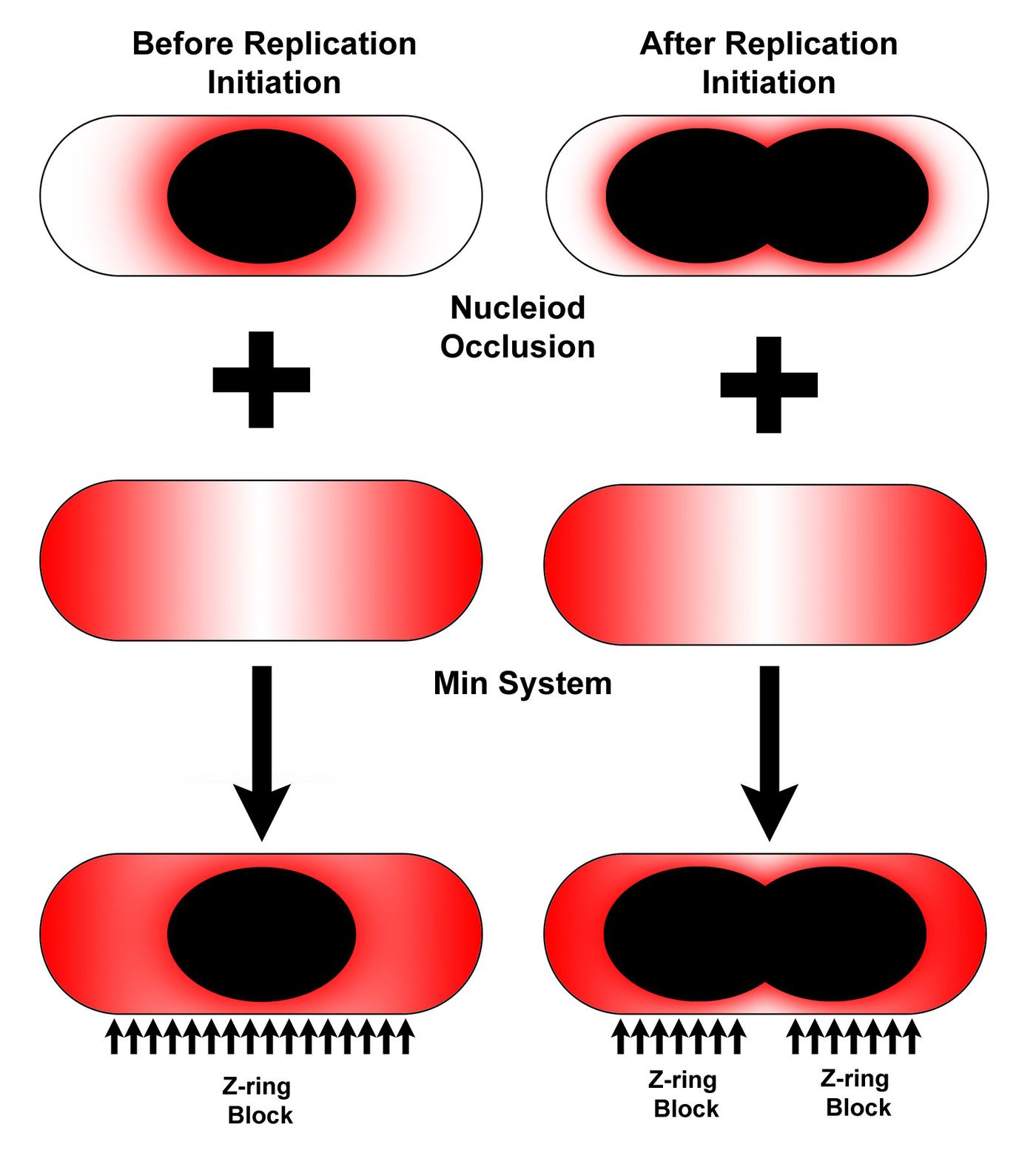
Figure 1.1: A pictorial representation of the coordinated effects of NO and the Min system on division site selection. In all cases, red shading represents a block to Z-ring assembly. The left column corresponds to the scenario before significant chromosome replication. NO and Min block division at all sites along the length of the cell. In the right column, significant clearing of the division block from the midcell region is apparent after the chromosomes begin to segregate.
1.2.2. The division site marker “FtsZ”
The bacterial cell division genes have largely been identified by the isolation of conditional mutants that, at elevated temperatures, fail to divide and instead form long filamentous structures. The first such fts (filamentation temperature sensitive) mutant to be identified was ftsZ (Lutkenhaus, Wolf-Watz et al. 1980), the gene for the FtsZ protein. ftsZ orthologs are found almost ubiquitously across eubacteria and archaebacteria and are also found in the organelles of several eukaryotic species, most notably in chloroplasts (where it plays a role in plastid division) and the mitochondria of some unicellular eukaryotes (Margolin 2000). While mitochondrial division in higher eukaryotes is typically performed by dynamin structures which operate from the outside of the organelle, the presence of FtsZ orthologs in the organelles of lower eukaryotes is in line with the hypothesis that organelles originated as symbiotic inclusions into the eukaryotic cytoplasm. Surprisingly, FtsZ homologues are absent from several eubacterial species, including the Chlamydiae family (Margolin 2000), This obligate intracellular pathogen may, however, utilize elements from the cytoplasm of its eukaryotic host for division. It is also absent from the archea bacterium Aeropyrum pernix and Ureaplasma urealyticum (Margolin 2000). Whether these organisms have a divergent, yet functionally similar, protein to perform the role of FtsZ is unknown.
1.2.2.1 The role of FtsZ and the Z-ring in cell division
It has been established that FtsZ is the essential initiator of divisome assembly in both E. coli (Dai and Lutkenhaus 1991) and B. subtilis (Beall and Lutkenhaus 1991). FtsZ was subsequently shown to form a ring (the Z-ring) at the nascent division site by immunoelectronmicroscopy in E. coli (Bi and Lutkenhaus 1991). Several years later a similar circumferential ring of FtsZ protein was observed in B. subtilis for both medial and polar (sporulation associated) division events (Levin and Losick 1996), this time utilizing fluorescent microscopy with GFP-tagged FtsZ. The presence of a discernable ring of protein suggests the presence of a cytoskeletal structure in bacteria. The Z-ring contracts along with membrane invagination during division (Bi and Lutkenhaus 1991). Once a Z-ring has assembled, it becomes competent to recruit other division proteins to the division site (see sections 1.2.2.3 and 1.2.3). As yet no other protein or factor has been identified that locates to the division site before the Z-ring forms. All other proteins that assemble at the division site require prior formation of the Z-ring to localize (reviewed in detail in (Errington, Daniel et al. 2003)).
1.2.2.2 The biochemistry of FtsZ
The FtsZ protein monomer is approximately 40 kDa in size. While there is little primary structure homology between FtsZ and the eukaryotic cytoskeletal protein tubulin (~10%) these two GTPases have similar tertiary structures (Lowe and Amos 1998; Nogales, Wolf et al. 1998)(see also Figure 1.2A).
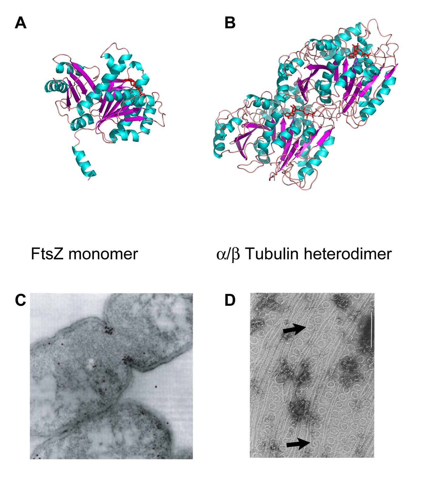
Figure 1.2: (A) X-ray crystal structure of FtsZ with bound GDP (in red). (B) X-ray structure of the / tubulin heterodimer (bound GTP is in red). (C) Electron micrograph of immunogold labeled FtsZ in E. coli. Electron-dark areas at the division furrow demonstrate the presence of FtsZ at the division site. (D) Electron micrograph of polymerized FtsZ in the presence of GDP. Protofilaments, sheets and minirings (arrows) can be visualized (bar = 100 m).
FtsZ binds GTP and subsequently polymerizes into linear protofilaments of only one molecule in width (Mukherjee and Lutkenhaus 1994). This contrasts with the tubulin protofilament, which is composed of a heterodimer of two highly homologous and subunits which form protofilaments two molecules wide. These FtsZ protofilaments can ultimately form two-dimensional sheets (Erickson, Taylor et al. 1996). In the presence of GDP or, presumably, upon GTP hydrolysis and release of inorganic phosphate (Pi), protofilaments can adopt a circular conformation with a diameter of 15-25 nm (Erickson, Taylor et al. 1996; Lu, Reedy et al. 2000). This diameter (and hence curvature) is significantly smaller (tighter) than the width of a typical eubacterial cell. Consequently, it is possible that hydrolysis of GTP and release of Pi result in an arced filament that, if attached to the membrane, would cause a local invagination (see Section 1.2.4.1 for further discussion).
Polymerization permits hydrolysis of the bound GTP molecule, although whether straight protofilaments are composed of FtsZ bound to GTP, or FtsZ bound to GDP + Pi, is controversial (Mingorance, Rueda et al. 2001; Scheffers and Driessen 2002; Romberg and Mitchison 2004). The debate has relevance to the action of FtsZ filaments and their involvement in creating the force of division. As Romberg and Levin (2003) point out, a polymer of FtsZ-GDP has stored within it the energy of many -phosphate hydrolysis events (see Section 1.2.4.1 for further discussion on the action of FtsZ filaments during division). They argue that this “spring-loaded” hypothesis is more likely than a model of sequential or simultaneous hydrolysis upon a signal to divide, although, the rate at which Z-ring needs to contract during division and the sort of forces it is required to exert are currently unknown.
Two well-characterized mutants of FtsZ in E. coli have shed light on FtsZ function. The ftsZ84 mutant, first described 25 years ago (Lutkenhaus, Wolf-Watz et al. 1980), results from a single point mutation that changes Gly105 to a serine (numbering refers to the E. coli protein). When a strain carrying ftsZ84 is shifted to the non-permissive temperature (42 ºC) Z-rings disappear within 1 minute (Addinall, Cao et al. 1997). Gly105 is involved in GTP binding (Nogales, Downing et al. 1998) and the mutation to serine slows rate of GTP hydrolysis. Since GTP binding and hydrolysis involve contacts between opposite faces of two FtsZ molecules (Nogales, Downing et al. 1998) the temperature-sensitive depolymerization phenomenon is most probably the result of loss of binding of GTP, rather than rapid hydrolysis of GTP to the more weakly polymerized GDP form. Interestingly the temperature-sensitive phenotype of this mutant can be recovered with high salt. It is possible that the ftsZ84 mutation results in a poorly “spring-loaded” filament due to an inability to form a strong polymer in the GDP + Pi form, preferring to depolymerize rather than exert a force on the membrane. A high salt concentration in the media may lessen the force required by balancing the osmotic pressure differentials between inside and outside of the cell, allowing this weaker filament to support division at the non-permissive temperature (see section 1.2.4.1 for further discussion).
The second well-characterized mutation in E. coli FtsZ is the ftsZ26 allele. During a screen for FtsZ mutants, Bi and Lutkenhaus (1992) isolated a strain in which cell division is blocked at the non-permissive temperature. In addition, even at the permissive temperature, division site morphology was frequently not planar. Cell wall and membrane invagination were skewed away from the medial plane as well, and the cells formed “screw-like” invaginations. That is, the invaginations observed appear to form as a coil with a large pitch. These invaginations correlate with FtsZ protein as shown by immunoelectron microscopy (Bi and Lutkenhaus 1992) and were observed more clearly (in 3-dimensions) by scanning electron microscopy (Addinall and Lutkenhaus 1996). This result is intriguing as it suggests: (i) FtsZ polymerization can take place over a considerable interval of space at mid-cell, yet under normal conditions the ring forms precisely at mid-cell and; (ii) invagination of the membrane and cell wall is very precisely localized to the Z-ring, even when the Z-ring is distorted. Why the Z-ring becomes distorted into a coil with a large pitch is an intriguing and unanswered question. It is also remarkable that these large-pitched Z-rings can still support membrane and cell wall invagination. It is tempting to speculate that normal Z-rings may also contain a coiled structure however with much smaller pitch, which gives the appearance of being a true ring. The potential coiled nature of Z-rings and their impact on Z-ring constriction is discussed further in Section 1.2.4.1.
1.2.2.3 FtsZ stabilization and dynamics
Several proteins that bind FtsZ modulate Z-ring formation directly. Many of these proteins have functionally similar roles to proteins that bind and decorate eukaryotic tubulin, however there is no primary structure homology between eukaryotic and prokaryotic modulators (Romberg and Levin 2003; Gitai 2005; Goehring and Beckwith 2005). In addition to the activity of MinC discussed above (Section 1.2.2) the following proteins also play a role in FtsZ dynamics and stabilization.
EzrA: EzrA is a Z-ring associated protein that is typically found only in low-GC Gram-positive bacteria including B. subtilis. The ezrA gene was isolated in a screen for lethality when combined with minCD in B. subtilis (Levin, Kurtser et al. 1999). EzrA contains a single transmembrane domain and a larger cytoplasmic domain and it localizes diffusely to the membrane with some concentration at the site of Z-rings. In the absence of EzrA, Z-rings have the ability to form near cell poles, much like in a minCD mutant, implying that MinCD alone is not sufficient to prevent the formation of Z-rings at the cell poles of B. subtilis. The somewhat paradoxical concentration of this Z-ring destabilizer at a formed Z-ring suggests that EzrA can not overcome stabilizing forces when the Z-ring is appropriately formed and positioned but it remains associated with it nonetheless. The nature of the interaction with the Z-ring is entirely unknown.
ZipA: ZipA is another protein with a single transmembrane span and a large cytoplasmic domain (Hale and de Boer 1997). ZipA, however, is found only in enteric Gram negative bacteria like E. coli. ZipA apparently stabilizes the Z-ring as overexpression of ZipA can compensate for the temperature-sensitive ftsZ84 phenotype (RayChaudhuri 1999). In vitro experiments with cytoplasmic ZipA and FtsZ have shown that ZipA enhances bundling of FtsZ protofilaments (RayChaudhuri 1999). The presence of a transmembrane domain in ZipA suggests that the protein plays a role in tethering Z-rings to the membrane and/or transmitting force from the Z-ring to the membrane during invagination. However, if this is the role of ZipA, the absence of the protein from many bacterial species leaves open the question as to how these bacteria might apply the force of the Z-ring to the membrane. One possibility is that this role is assumed by the much more widely conserved FtsA protein.
ZapA: ZapA was discovered due to its ability to augment Z-ring structures in the presence of MinD overexpression (Gueiros-Filho and Losick 2002), an excess of which typically inhibits cell division by preventing stable Z-rings from forming (see Section 1.2.1.2). ZapA is a small, 10 kDa protein with no apparent membrane-anchoring or transmembrane sequence. ZapA orthologous are ubiquitous among bacterial species. ZapA co-localizes with FtsZ in B. subtilis and E. coli, suggesting it is part of the cytokinetic machinery. When a zapA null is combined with a divIVA null (the phenotype of which is described in Section 1.2.1.2) the division block is even further enhanced (Gueiros-Filho and Losick 2002). This is congruent with the notion that since MinCD is free to block division everywhere in the absence of DivIVA, the absence of ZapA results in even fewer Z-ring formations. Then again, a zapA deletion combined with an ezrA deletion also leads to a division block (Gueiros-Filho and Losick 2002). This is surprising as ZapA would appear to be a positive regulator of Z-rings yet EzrA is a negative regulator (see above). Clearly, the molecular basis by which each of these proteins regulates Z-ring formation is not fully understood.
It is known that ZapA can enhance FtsZ protofilament bundling in vitro (Gueiros-Filho and Losick 2002) and that it probably does so by forming parallel dimers with each ZapA monomer capable of binding a single protofilament (Low, Moncrieffe et al. 2004). The crystal structure of ZapA from Pseudomonas aeruginosa has been determined (Low, Moncrieffe et al. 2004) revealing an elongated structure composed of a globular N-terminal domain followed by a C-terminal coiled coil. In the crystal, there were close contacts between two monomers in the C-terminal coiled-coil region, suggesting a dimeric structure, and dimers have been observed in solution (Gueiros-Filho and Losick 2002). It has been suggested that the N-terminal globular domain is the site for interactions with FtsZ filaments (A. Handler, F. Gueiros-Filho, and R. Losick, personal communication).
FtsA: FtsA has primary structure homology to the actin family of proteins (Bork, Sander et al. 1992) and the crystal structure revealed a 3D fold also typical of the actin family (van den Ent and Lowe 2000). Despite similarities to actin, FtsA has not been observed to polymerize in vitro. In the case of E. coli this is not too surprising, as the ratio of FtsZ to FtsA is 100:1 (Dai and Lutkenhaus 1992), thus there does not appear to be sufficient FtsA to form functionally relevant filaments. In B. subtilis the ratio of FtsZ to FtsA is approximately 5:1 (Feucht, Lucet et al. 2001) suggesting the extra abundance of FtsA may be sufficient to form functionally relevant actin-like filaments. B. subtilis FtsA has been observed to dimerize and hydrolyze ATP in vitro (Feucht, Lucet et al. 2001), however no actin-like filament formation has been observed. The persistence of ATPase activity, despite the protein’s apparent inability to form filaments, suggests that ATP hydrolysis is essential for the function of FtsA. The effect of direct interruption of the ATP binding pocket of FtsA has not yet been assessed. FtsA appears to be associated with cell membranes (Sanchez, Valencia et al. 1994) and, as an FtsZ binding protein, it may play a role in targeting FtsZ to the membrane. More recently it was shown that FtsA contains a short C-terminal amphipathic helix that directly targets it to the membrane (Pichoff and Lutkenhaus 2005). FtsA trafficking of FtsZ towards the cytoplasmic membrane may be at least partly responsible for generating sufficient concentration of FtsZ molecules to allow polymerization to take place preferentially at the membrane. It may also play a role in FtsZ subunit turnover, returning FtsZ molecules from the cytoplasm to the Z-ring structure (see FtsZ dynamics below).
FtsZ dynamics: Studies of Z-ring assembly and disassembly have established that these processes can occur relatively rapidly on the time scale of 1 minute (Addinall, Cao et al. 1997; Sun and Margolin 1998; Rueda, Vicente et al. 2003). Amazingly, this rapid assembly/disassembly is also accompanied by another dynamic behavior — rapid exchange between FtsZ monomers in the cytoplasm and a static Z-ring. Utilizing FRAP (Flourescence Recovery After Photobleaching). The half-time for remodeling of the E. coli Z-ring from monomers in solution was measured at approximately 30 seconds (Stricker, Maddox et al. 2002). A more recent study in E. coli and B. subtilis indicated that the half-time is closer to 9 seconds (Anderson, Gueiros-Filho et al. 2004). This more recent measure of hydrolysis rate is more in line with the GTP hydrolysis rate of FtsZ (5-10 GTP per FtsZ per minute) in protofilaments (Romberg and Mitchison 2004)
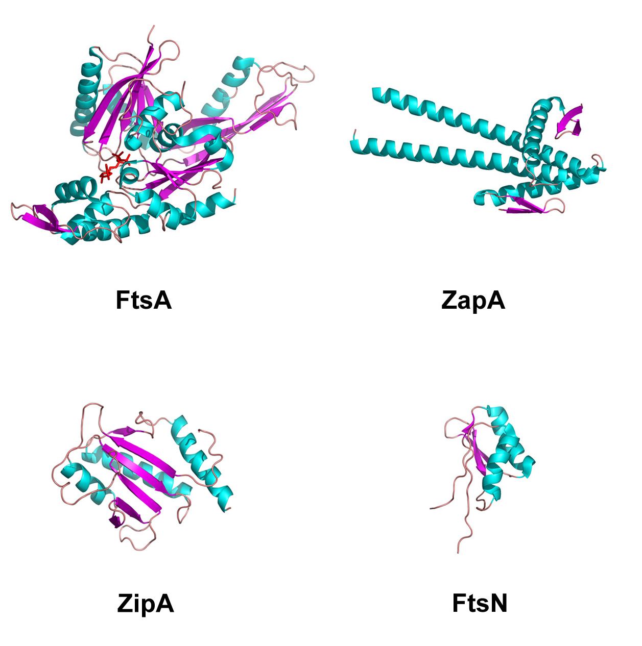
Figure 1.3: Structures of divisomal proteins. Apart from FtsZ, there are only four divisomal proteins whose structures have been determined. The FtsA structure definitively places it amongst the actin family of proteins. The crystal structure of ZapA reveals a homodimer which may in part explain its ability to enhance bundling of FtsZ protofilaments. The C-terminal domain of ZipA has revealed how it interacts with FtsZ. The NMR structure of FtsN is reminiscent of the RNA binding fold also found in murein binding proteins.
and suggests that GTP hydrolysis is the rate limiting step in FtsZ monomer turnover. In line with this hypothesis is the observation that the FtsZ mutant ftsZ84, which hydrolyses GTP at a slower rate than wildtype FtsZ, undergoes exchange between its Z-ring and monomer forms at a slower rate than does wildtype FtsZ (Anderson, Gueiros-Filho et al. 2004). Experiments in which ZapA, EzrA, and MinCD were manipulated showed that these proteins had no effect on the turnover rate (Anderson, Gueiros-Filho et al. 2004), implying that these proteins may only play a role in bundling of FtsZ protofilaments, and not in FtsZ monomer turnover.
FtsZ has also been observed to undergo macromolecular movements. Ben-Yehuda and Losick (2002), using fluorescence microscopy on B. subtilis cells, reported the movement of medial Z-rings to polar locations (under conditions that produce sporulation and hence division at pole sites) via a coiled or helical structure with a large pitch (inaccurately referred to as a spiral structure both in this paper and elsewhere). Remarkably, when cells are induced back into a vegetative growth pattern, Z-rings returned to a medial location via the same large-pitched coiled structure. Z-ring coils have been noted before in the literature. As mentioned above, the ftsZ26 mutant apparently can form a coiled structure with large pitch spontaneously. Simple overexpression of FtsZ in E. coli gives rise to coiled structures as well (Ma, Ehrhardt et al. 1996). E. coli cells that have been engineered to be devoid of phosphatidylethanolamine also contain large-pitched, coil-like Z-ring structures (Mileykovskaya, Sun et al. 1998) which suggests membrane interactions or dynamics might be, in part, responsible for supporting highly pitched Z-rings.
These highly pitched Z-ring structures may represent an alternative but functionally relevant form for the Z-ring. Under standard conditions the Z-ring would not actually be a true ring, but rather a very tight (small pitched) coil. Each loop of the coil would stably interact with its neighbor, as FtsZ protofilaments are known to form stable sheets. This bundling is modulated by the proteins listed above and by several more including the SulA protein of the SOS response in E. coli (Trusca, Scott et al. 1998) and SpoIIE during sporulation in B. subtilis (Ben-Yehuda and Losick 2002). However, the ability to form a more highly pitched coil as the result of membrane changes or modulating proteins may aid in either the disassembly of the ring or the trafficking of it to polar regions for division events there.
1.2.3 Assembly of the Divisome
The above discussion suggests that Z-ring maintenance is a complicated process that involves several proteins beyond polymerized FtsZ. These proteins play important roles in regulating Z-ring formation in space and time and as such can be considered as part of a “Z-ring network” of proteins. Subsequent to the establishment of the Z-ring, several other proteins have been noted to localize to the incipient division site and, at least in E. coli, there appears to be a hierarchical assembly of these proteins (Goehring and Beckwith 2005) (see figure 1.4). That is, assembly of one protein type at midcell is followed by the assembly at midcell of the next protein type and so forth.
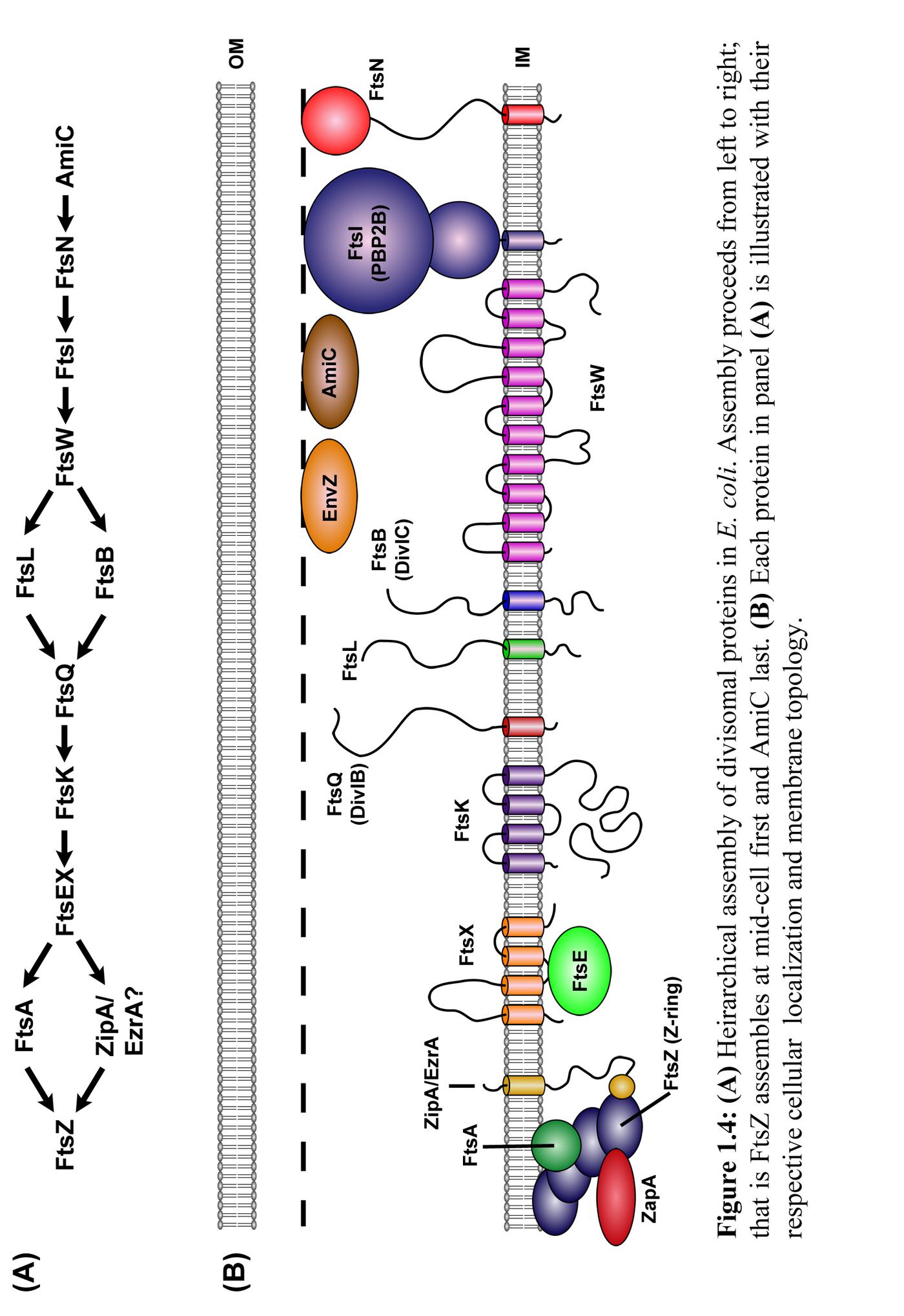
This section describes the major proteins that localize to the midcell after Z-ring formation. These proteins have largely been identified by the generation of fts- alleles in E. coli and B. subtilis. They appear to operate cooperatively as deletion or depletion of any of these proteins result in division arrest. Each of the major proteins will be briefly introduced in the order with which they are believed to assemble in E. coli (see Ffigure 1.4), however it should be noted that this order is still currently being tested experimentally. It should also be noted that as of this writing our understanding of how these proteins interact with each other or the Z-ring id rudimentary at best. It can not be assumed that because one protein follows another in the divisomal network that it directly or even indirectly interacts with the protein preceding it.
1.2.3.1 FtsEX
The genes ftsE and ftsX are widely conserved in eubacteria. Deletion of these genes in E. coli is not lethal, but rather results in the generation of a filamentous phenotype. Intriguingly, this phenotype partially reverts in the presence of high salt (de Leeuw, Graham et al. 1999; Schmidt, Peterson et al. 2004). The protein products of these two genes form a complex (FtsEX) which has strong homology to the ABC-type transport systems, with FtsE comprising the ATP-binding module and FtsX comprising the membrane pore (de Leeuw, Graham et al. 1999; Bernatchez, Francis et al. 2000). Sequence comparisons between FtsEX and other ABC-transporters suggest FtsEX groups best with importers rather than exporters (Schmidt, Peterson et al. 2004), although it is yet to be established what, if anything, FtsEX transports. Localization studies of FtsEX in E. coli have determined that this complex arrives at the division site after assembly of the FtsZ ring and requires FtsA and ZipA for localization as well. FtsK, FtsQ, and FtsL, however, fail to localize in the absence of FtsEX (Schmidt, Peterson et al. 2004) (see Figure 1.4A). There are homologs of ftsE and ftsX in B. subtilis, but their roles in cell division have not been examined. Aberrant DNA condensation has also been noted in mutant ftsE strains of Neisseria gonorrhoeae (Bernatchez, Francis et al. 2000), suggesting the FtsEX complex may be involved in chromosomal preparation for division.
1.2.3.2. FtsK
FtsK is a large multi-domain protein comprising a large C-terminal cytoplasmic domain and a smaller N-terminal domain consisting of four membrane spans; the membrane portion targets the protein to the division site (Wang and Lutkenhaus 1998). The C-terminal domain is an ATP dependant DNA translocase which appears to be responsible for resolution of chromosomal dimers (Steiner, Liu et al. 1999). FtsK may act as a checkpoint that ensures unsegregated chromosomes are not severed by a closing division septum (Rothfield, Justice et al. 1999). The proximal localization of FtsEX (and its apparent association with DNA condensation) may ultimately be tied to this process of chromosomal segregation.
1.2.3.3. DivIB/FtsQ
E. coli FtsQ and the B. subtilis ortholog DivIB have a similar bitopic topology with a small cytoplasmic domain, a single transmembrane domain and a larger extracytoplasmic domain. Despite both DivIB and FtsQ localizing to mid-cell during division (Harry and Wake 1997; Buddelmeijer, Aarsman et al. 1998) their role in cell division remains enigmatic. Interestingly, the relative abundance of these proteins in their respective hosts differs greatly. FtsQ is essential in E. coli and yet has an extremely low abundance at approximately 22 molecules per cell (Carson, Barondess et al. 1991). In contrast, B. subtilis DivIB is highly abundant at approximately 5,000–13,000 copies per cell (Rowland, Katis et al. 1997) and yet is only essential at growth temperatures in excess of 48 ºC(Beall and Lutkenhaus 1989; Harry and Wake 1989). At permissive temperatures (down to 30 ºC), cell filamentation occurs in a divIB null, but cells continue to grow. Only the extracytoplasmic domain appears to be functional as the short cytoplasmic domain and the transmembrane domains of these proteins can be replaced without loss of function (Guzman, Weiss et al. 1997; Buddelmeijer, Aarsman et al. 1998; Katis and Wake 1999). Interestingly apart from premature truncation of the polypeptide chain, all mutant alleles of DivIB/FtsQ are localized in the extreme C-terminal region (Harry, Stewart et al. 1993; Chen, Minev et al. 2002) suggesting this region is crucial for function. Several lines of circumstantial evidence imply that DivIB/FtsQ plays a role in peptidoglycan synthesis or remodeling: (i) wall-less bacteria are apparently missing orthologs of DivIB/FtsQ; and (ii) the greater abundance of DivIB over FtsQ correlates well with the need for B. subtilis to construct a thicker wall of peptidoglycan than E. coli; (iii) FtsQ from E. coli has low sequence homology to Mpl (~17%), an enzyme involved in peptidoglycan recycling (Chen, Minev et al. 2002). The DivIB/FtsQ family of proteins have, however, not been ascribed any enzymatic activity to date. FtsQ requires the localization of FtsK before it can localize to the division site in E. coli (Chen and Beckwith 2001), however the dependence of B. subtilis DivIB localization on FtsK has not been determined.
1.2.3.4 FtsL
The ftsL gene was first identified as an essential division gene in E. coli (Guzman, Barondess et al. 1992) and later identified in B. subtilis (Daniel, Williams et al. 1996). FtsL is a small protein of approximately 14 kDa, comprising a short cytoplasmic domain, a single transmembrane segment and a larger extracytoplasmic domain that contains a predicted coiled-coil region proximal to the membrane (Guzman, Barondess et al. 1992; Daniel, Harry et al. 1998). In B. subtilis, depletion of FtsL inhibits cell division and results in the formation of filamentous cells with no apparent septation (no partial constrictions). B. subtilis ftsL appears to be highly malleable as B. licheniformis, B. badius and B. circulans ftsL can complement an ftsL null in B. subtilis despite B. badius ftsL and B. circulans ftsL having lower than 40% identity with B. subtilis ftsL (Sievers and Errington 2000). In addition, the putative coiled-coil region can be extensively mutagenized (even at the crucial ‘d’ positions that define the interhelical interface a coiled-coil) and still remain functional (Sievers and Errington 2000). Nonetheless, deletion of the last 10 C-terminal residues eliminates function (Sievers and Errington 2000).
1.2.3.5. DivIC/FtsB
The divIC gene was first identified in B. subtilis and shown to be an essential division gene (Levin and Losick 1994). DivIC is a similar size to FtsL and also has a similar bitopic topology including a predicted coiled-coil region proximal to the membrane. In addition, the extreme C-terminal residues appear to be functionally important, as a five base-pair duplication in this region that results a frame-shift and an increase in the length of the protein by seven residues causes a temperature-sensitive phenotype (the div-355 mutant). Only the extracytoplasmic region is essential for function, as a divIC gene in which the cytoplasmic and transmembrane have been replaced with domains from the unrelated TolR protein is still functional (Katis and Wake 1999). A potential E. coli ortholog of DivIC (first identified as the ygbQ open reading frame and then renamed as ftsB) was recently identified and shown to be essential for division in E. coli and Vibrio cholerae (Buddelmeijer, Judson et al. 2002). FtsB has a similar membrane topology and localizes to midcell during division but has only 14% sequence homology to B. subtilis DivIC. (Buddelmeijer, Judson et al. 2002). Whether FtsB is a true ortholog of DivIC in E. coli is not clear (especially since it shares little sequence homology) but its similar size, membrane topology, cell division phenotype, midcell localization and the fact it is located distally from other cell division genes on the chromosome–all properties it shares with B. subtilis DivIC–suggests it is likely to be an ortholog.
1.2.3.6. FtsW
First cloned and sequenced from E. coli in 1989, FtsW is a large integral membrane protein with high sequence homology to E. coli RodA (32% identical) and to B. subtilis SpoVE (40% identical) (Ikeda, Sato et al. 1989). These three proteins have been classed in the SEDS (shape, elongation, division, and sporulation) family of polytopic proteins (Henriques, Glaser et al. 1998). Each SEDS protein appears to be functionally linked to a specific penicillin binding protein (PBP). For example, in E. coli, RodA is linked with cylindrical peptidoglycan synthesis via PBP2, B. subtilis SpoVE is linked with SpoVD, a spore cortex synthetic enzyme, while FtsW appears to be associated with FtsI for the septal synthesis of septal murein (see Section 1.2.3.8). Because FtsW is a large, polytopic, membrane protein, it has been suggested that it may be involved with the translocation or “flipping” of the peptidoglycan precursor, lipid II (Ehlert and Holtje 1996). In accordance with this hypothesis, there appears to be a perfect correlation between the presence of FtsW and FtsI in sequenced genomes to date (Mercer and Weiss 2002) and notably both are missing from wall-less bacteria (e.g. Mycoplasma genitalium) (Khattar, Addinall et al. 1997). It has also been shown that FtsW is recruited to the septum after FtsL and is required to recruit FtsI (Mercer and Weiss 2002), placing FtsW between FtsL and FtsI on the hierarchical assembly line. It has been known for some time that B. subtilis contains an FtsW-like homolog (the ylaO ORF) (Henriques, Glaser et al. 1998) and unpublished experimental results show that this protein localizes to midcell (Errington, Daniel et al. 2003). Interestingly, while FtsW and FtsI are both located in the mra cluster of cell division and peptidoglycan synthesis genes in E. coli (Ikeda, Sato et al. 1989), FtsW is located some 30 kilobases upstream of PBP2B (the ortholog of FtsI) in B. subtilis.
1.2.3.7. PBP2B/FtsI
B. subtilis PBP2B and the E. coli ortholog FtsI are both bitopic class B penicillin binding proteins (PBPs) with a short cytoplasmic region, a single transmembrane segment and a much larger extracytoplasmic region. This extracytoplasmic region has been shown to consist of two domains: an N-terminal non-catalytic domain followed by a PBP with transpeptidase activity (Nguyen-Disteche, Fraipont et al. 1998). This is in contrast to class A PBPs which contain both transpeptidase and transglycosylase activity. Because class B PBPs contain only a transpeptidase activity they are unable to lengthen peptidoglycan layers and presumably function by cross-linking layers instead (Holtje 1998). Several lines of evidence suggest the N-terminal non-catalytic domain mediates the localization of FtsI to mid-cell and its interactions with other divisomal proteins (Marrec-Fairley, Piette et al. 2000; Wissel and Weiss 2004), however more recently is was shown that the transmembrane segment is sufficient to target GFP (and presumably the entire FtsI molecule) to the mid-cell. In B. subtilis, PBP2B is accompanied at the division site by PBP1, a class A PBP with both transpeptidase and transglycosylase activity.
1.2.3.8. FtsN
FtsN was first isolated as a multicopy suppressor of a temperature-sensitive ftsA mutation in E. coli (Dai, Xu et al. 1993). Somewhat surprisingly, the multicopy FtsN mutant also suppressed, although to a lesser extent, temperature-sensitive mutations in ftsI, ftsQ, and ftsK. The gene sequence is poorly conserved in species with a clear homolog (largely enteric bacteria) and is not present in B. subtilis (Dai, Xu et al. 1996). FtsN is another bitopic protein with a small cytoplasmic region, a single transmembrane segment and a larger extracytoplasmic region. This region of FtsN is largely unstructured except for a small globular domain at the C-terminus for which the structure has been determined (Yang, Van Den Ent et al. 2004). This domain has 30% sequence identity with the murein-binding domain of B. subtilis CwlC, an N-acetylmuramoyl-L-amidase that most likely confers the observed ability of FtsN with its ability to bind murein sacculi from dividing E. coli cells (Ursinus, van den Ent et al. 2004). Binding however is reduced when murein is prepared from cells with division blocks or cells that lack the AmiA/B/C family of amidases that are responsible for the cleavage of the peptide cross-links in murein (see Section 1.2.4.3); this suggests that FtsN can identify a specific form of murein.
1.2.4. Membrane and Cell Wall Constriction and Cell Separation
While the assembly of the above components of the divisome is necessary for successful division to take place, it has not been established that assembly alone is sufficient. Indeed, the exact nature of the “signal” that triggers division upon assembly of the divisome is presently unknown. The physical act of division involves constriction of the membrane which apparently follows the constriction of the Z-ring itself; this is coordinated with the construction of cell wall between the newly formed cells.
1.2.4.1 Mechanisms of Z-ring constriction and membrane invagination
As division takes place, the Z-ring constricts into a smaller and smaller annulus, apparently moving the cytoplasmic membrane with it (Bi and Lutkenhaus 1991). The integrity of the ring is maintained in spite of shrinking in size. This is presumably necessary to preserve the appropriate localization of other divisomal proteins associated with it. Studies using the temperature-sensitive (ts) allele ftsZ84 indicate that disruption of Z-ring formation by temperature shifting during septation results in partial invagination. This demonstrates that the ring is essential throughout (Addinall, Cao et al. 1997); that is, the rest of the division apparatus will functionally arrest in the absence of the Z-ring. In turn, shifting of ftsZ84 cells to the permissive temperature is not sufficient to allow reassembly of the ring at a partially invaginated site. Thus, recruitment and/or activation of other division proteins by a Z-ring requires Z-ring formation at non-invaginated locations (Addinall, Cao et al. 1997). Similar temperature-shift experiments in E. coli cells using ts alleles of divisomal proteins that localize after FtsZ still have a slight invagination although division can not be completed (Taschner, Huls et al. 1988) suggesting that initiation of Z-ring constriction and completion of division can be uncoupled.
Given the structure of FtsZ (both tertiary and quaternary) and its similarities with tubulin it is tempting to suggest that Z-ring constriction results from analogous motive mechanisms employed by eukaryotic tubulins. A model of a tightly pitched helical Z-ring further suggests a mechanism in which FtsZ protofilaments are able to move against each other in a similar mechanism as in the actomyosin ring in eukaryotes (Bramhill and Thompson 1994). In this model, movement of FtsZ protofilaments against each other would tighten the coil, reducing the aperture of the annulus. However, no “motor” proteins typically associated with movement of tubulin in eukaryotes have ever been discovered in the prokaryotic world. Tightening of the coil has also been suggested to result from hydrolysis of bound GTP to GDP in the FtsZ filaments. This hydrolysis event and/or the release of Pi from filaments leads to a conformational change, in effect bending filaments into a tighter arc (Lu, Reedy et al. 2000). Both these hypotheses rely on FtsZ filaments being tightly tethered to the cell membrane. As discussed above, FtsZ is attached to the membrane via FtsA, ZipA (in E. coli) and potentially EzrA (in B. subtilis), although whether these interactions are sufficiently strong to pull on the cytoplasmic membrane is unknown. It is also tempting to hypothesize that constriction is, at least in part, directed by construction of cell wall in the septal space during division. Wall-less bacteria (e.g. the mycoplasma family) however, are able to achieve division without cell wall synthesis, indicating that synthesis of cell wall material is not always essential for initiation or completion of division.
1.2.4.2 Synthesis of Cell Wall in the Septal Space
It would appear that Z-ring constriction and completed membrane invagination can be uncoupled from the synthesis of cell wall in the septal space between the two newly formed cells, at least in B. subtilis. Depletion of the divisomal transpeptidase PBP2B (FtsI in E. coli) leads to a division block, yet Z-rings still form regularly while DivIB, FtsL and DivIC do not localize (Daniel, Harry et al. 2000). Remarkably, complete membrane invaginations could be seen at some sites without any associated septal peptidoglycan synthesis. Two major conclusions were drawn from this study: (i) DivIB, FtsL, and DivIC are likely to be involved in murein synthesis (and not just remodeling) in the septal space; and (ii) some residual PBP2B activity after depletion may be sufficient to synthesize enough “appropriate” murein to initiate Z-ring constriction and membrane invagination; this is sufficient to trigger the invagination process, but is not sufficient to synthesize murein in the septal space.
In B. subtilis, PBP2B is accompanied at the divisome by another peptidoglycan synthesis enzyme, PBP1 (Pedersen, Angert et al. 1999). PBP1 belongs to the class A high-molecular-weight (HMW) group of PBPs which are capable of fully synthesizing peptidoglycan; that is they contain both transglycosylase and transpeptidase activity. Remarkably, deletion of the ponA gene, which encodes for PBP1, leads to aberrant Z-ring structures and improper localization (Pedersen, Angert et al. 1999). This in turn suggests a link between maintenance of the Z-ring and peptidoglycan synthesis at the division site.
1.2.4.3 Cell Separation
Cell separation mutants have been known in Gram positive bacteria for almost four decades. These “chaining” mutants where shown to be deficient in murein hydrolyases (Tomasz 1968; Fan 1970). Presumably, successful separation of two daughter cells after division and septal peptidoglycan synthesis requires the lysis of cross-linked murein between the two new cells.
It was not until much more recently that similar hydrolyases where identified in E. coli with the discovery of several murein amidases (responsible for the cleavage of the peptide sidechain of murein from the poly-sugar backbone) that are necessary for cell separation. Heidrich et. al. (2001) described the presence of three such amidases, AmiA, AmiB and AmiC. Deletion of all three genes resulted in a chaining phenotype in which septation is complete for the inner membrane but not the outer membrane. They also showed that murein at these septation sites was preferentially digested by AmiC, suggesting a specific murein structure may be present at division sites. Later, it was shown that AmiA and AmiC are transported to the periplasm where they localize diffusely around the entire cell with AmiC also concentrating at septal rings in constricting cells (Bernhardt and de Boer 2003). Furthermore, this localization of AmiC requires FtsN, demonstrating that AmiC localizes to the divisome after FtsN. Bernhardt et. al. hypothesize that FtsN may function by changing the murein structure locally at the division site, allowing AmiC to preferentially localize there.
A further murein hydrolase, EnvC, has more recently been shown to localize to division sites (Bernhardt and de Boer 2004). Using an “in-gel” assay, they showed EnvC has murein hydrolase activitiy. It has been known since the 1970s that mutations in this gene result in chaining mutants (Rodolakis, Thomas et al. 1973). Remarkably, Bernhardt and de Boer (2004), while searching for synthetic lethal mutants in combination with a deletion of the Min system, rediscovered the role of EnvC in cell division. The apparent link between the early acting Min system and the late acting EnvC murein hydrolase in the cell division process is notable and suggests that ultimately the entire process of division is essentially a completely coupled system.
1.3. DivIB, FtsL and DivIC: Three Enigmatic Proteins
The focus of this thesis is assembly of the bacterial divisome. As such, the primary focus of the experimental work has centered on the components of the divisome that remain the most enigmatic as far as their function and structure are concerned. Although all of the protein components of the divisome are necessary for successful division to take place, which in turn implies that the whole organization acts as a coordinated system to achieve this goal, it is clear that some components have specific roles in the process. For example, FtsZ is central to the formation of the division site at midcell, FtsI/PBP2B contains murein transpeptidase activity, and deletion of this gene in B. subtilis has a direct effect in septal murein synthesis. Indeed, the above discussion has implicated almost all of the components in “some aspect of the division process”, even though their precise functions are largely unknown. The three proteins, DivIB/FtsQ, FtsL and DivIC/FtsB (henceforth referred to as the BLC group) fail to be classified this way. While it is true that eubacteria that synthesize a cell wall tend to all have the BLC group, their link to murein synthesis is by this correlation only. FtsI/PBP2B, FtsN, and even FtsW have enzymatic, binding and structural associations that link them directly to murein synthesis. In contrast, the BLC group lacks any homology to the known proteins in the established synthetic pathways of peptidoglycan. What exactly can the BLC group be doing, if anything, that is related to murein synthesis and more generally during division as a whole? This study has focused on these three proteins exclusively in an attempt to answer this question.
1.3.1 Genomic Arrangment
The genomic arrangement of the BCL group has some intriguing properties, if inconclusive in how they relate to division. In both B. subtilis and E. coli, FtsL and DivIB/FtsQ are located close to one another (see Figure 1.5). This region of the chromosome has been known for some time to contain many genes related to cell division and murein synthesis (including most of the cytoplasmic enzymes responsible for peptidoglycan precursor synthesis and the well conserved ftsZ and ftsA genes) and has been coined the mra cluster (Ikeda, Wachi et al. 1990; Henriques, de Lencastre et al. 1992). The conservation of this genomic arrangement between these two very divergent species of eubacteria is highly suggestive of an ancient role for FtsL and DivIB in cellular division and murein synthesis.
In contrast, the genes for DivIC/FtsB lie in quite distal regions of the chromosome and are surrounded largely by genes of unknown function. The fact that these apparent orthologs are distally located in both B. subtilis and E. coli from the mra cluster is surprising. It is, however, a curious coincidence.
1.3.2 Dependency of Localization and Stability Studies
Much work on the BCL proteins has focused on their localization and stability in the absence of either one of the BCL group members or some other divisomal protein. Errington et al. (2003) recently reviewed the dependency of localization of various divisomal proteins. Table 1.1 summarizes these dependencies in B. subtilis. Upon depletion of DivIB – and at the nonpermissive temperature for a divIB null – FtsL is rapidly degraded (Daniel and Errington 2000) and DivIC becomes mislocalized (Katis, Wake et al. 2000); depletion of FtsL results in mislocalization of DivIC and DivIB (Daniel, Harry et al. 1998); and finally depletion of DivIC and FtsL leads to mislocalization of DivIB(Daniel and Errington 2000; Katis, Wake et al. 2000). It is impossible to account for all these results (without invoking unknown proteins), unless there is a co-operative assembly of the BCL proteins. This co-operativity extends to the PBP2B protein (Daniel, Harry et al. 2000) (see also Ttable 1.1). Unfortunately, there is an absence of localization data regarding the FtsEX and FtsK proteins, which, at least according to the E. coli assembly model (see Figure 1.4), precede assembly of the BLC
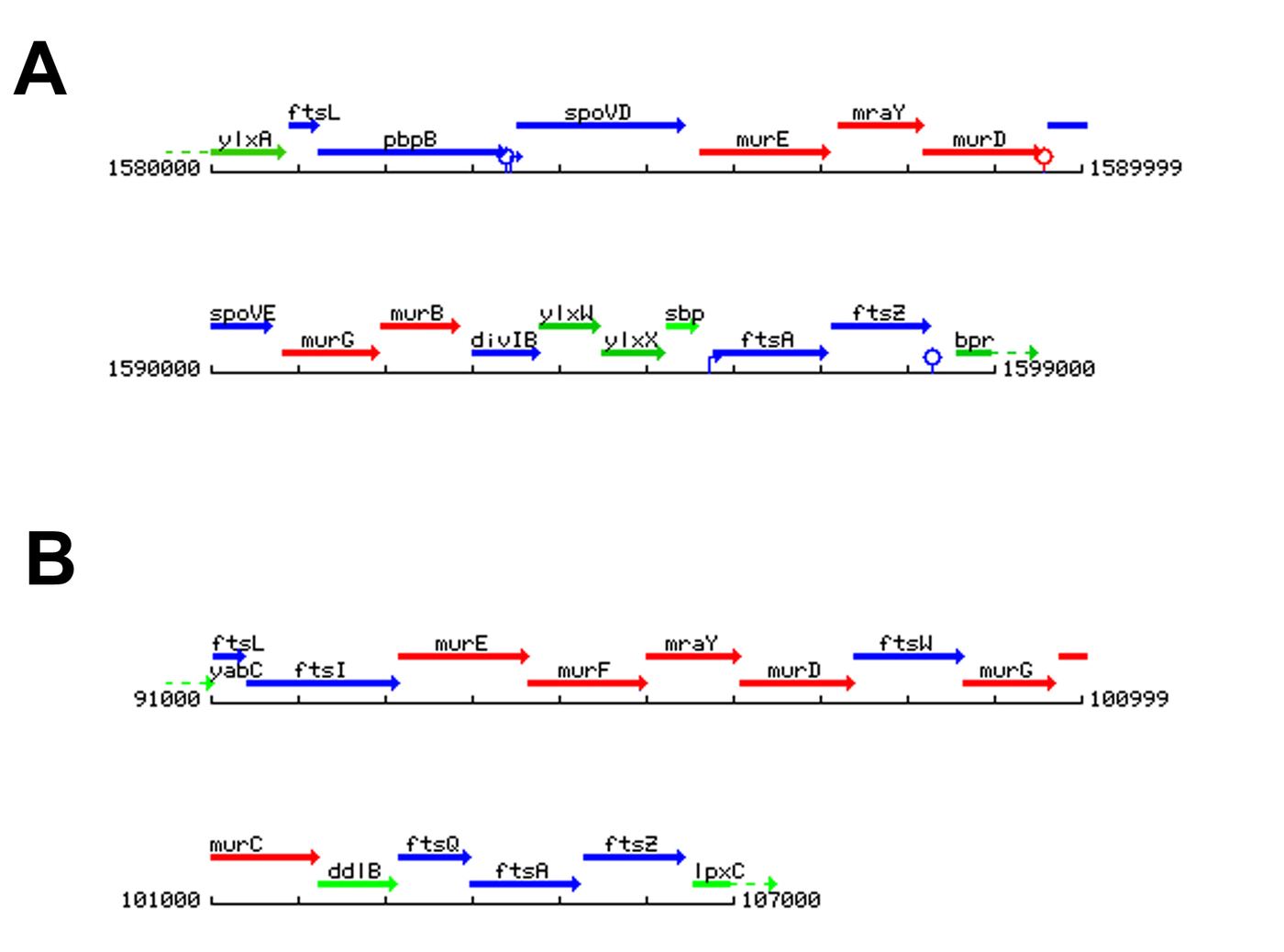
Figure 1.5: The genomic arrangement of cell division and cell wall synthesis genes (the mra cluster) in B. subtilis (A) and E. coli (B). Blue colored genes are related to cell division; red colored genes are related to peptidoglycan precursor synthesis and transport; green colored genes have either unknown functions or functions unrelated to either cell division or peptidoglycan metabolism. The numbers refer to base pairs in the genome. In both organisms, the genes that encode for DivIC and FtsB are located distal to the mra cluster. Figure adapted from http://genolist.pasteur.fr/SubtiList/ and http://genolist.pasteur.fr/Colibri/.
group. Interestingly, the linearity of assembly in E. coli is complicated by a presence of a co-operativity for assembly for FtsL and FtsB (Buddelmeijer, Judson et al. 2002) although it appears FtsQ is able to assemble in the absence of FtsL or FtsB in E. coli (Goehring, Gueiros-Filho et al. 2005).
Depletion of individual members of the BCL group not only has effects on localization, depletion can also affect the stability of other members. When a divIB null strain is placed at the non-permissive temperature, FtsL is rapidly degraded and there is an increase in the cellular level of DivIC (Katis, Wake et al. 2000). The increase in DivIC levels could stem from several phenomena, including, but not limited to, up-regulation because of filamentation at the nonpermissive temperature or aberrant degradation because of a lack of protease activity. Although FtsL and DivIC both contain coiled-coil domains, and there have been reports of a direct interaction between FtsL and DivIC (Sievers and Errington 2000), the disparity between the fates of FtsL and DivIC in a divIB null strain suggests otherwise. Depletion of FtsL results in an associated depletion of cellular levels of DivIC (Daniel, Harry et al. 1998). In contrast, Katis et al. (2000) showed that the decrease in FtsL observed in a divIB null strain coincides with an increase in DivIC levels. Since DivIB is present in the depletion study performed by Sievers et al. (2000), this gives indirect evidence that either DivIB directs, or DivIB is itself, a protease that degrades DivIC. Depletion of DivIC has no effect on FtsL levels (Daniel and Errington 2000).
| FtsZFtsZFtsZ | FtsAFtsAFtsA | DivIBDivIBDivIB | FtsLFtsLFtsL | DivICDivICDivIC | PBP2BPBP2BPBP2B | |
|---|---|---|---|---|---|---|
| FtsZFtsZFtsZ | nonono | nonono | nonono | n.d.n.d.n.d. | nonono | |
| FtsAFtsAFtsA | yesyesyes | nonono | n.d.n.d.n.d. | n.d.n.d.n.d. | n.d.n.d.n.d. | |
| DivIBDivIBDivIB | yesyesyes | n.d.n.d.n.d. | yesyesyes | yesyesyes | n.d.n.d.n.d. | |
| FtsLFtsLFtsL | yesyesyes | n.d.n.d.n.d. | yesyesyes | yesyesyes | yesyesyes | |
| DivICDivICDivIC | yesyesyes | yes*yes*yes* | yesyesyes | yesyesyes | yesyesyes | |
| PBP2BPBP2BPBP2B | yesyesyes | n.d.n.d.n.d. | yesyesyes | yesyesyes | yesyesyes |
Table 1.1: The dependency of localization of B. subtilis divisomal proteins on other divisomal proteins. A protein listed in the first column requires a protein listed in the first row for localization if the matching cross-cell indicates “yes”; e.g., all proteins in this table require FtsZ for localization (see second column). * stands for an unpublished result; n.d. stands for not determined. It can be seen that DivIB, FtsL and DivIC all require the presence of each other for localization. This co-operative assembly extends to the PBP2B protein, though the dependence of DivIB localization on PBP2B has not been determined. Table adapted from Errington et al. (2003).
1.3.3 The role of DivIB, FtsL and DivIC in Division.
The results discussed above seem to suggest that DivIB may directly stabilize FtsL, yet either directly or indirectly destabilize DivIC. As the stability of FtsL and DivIC do not correlate well, it is hard to envisage how they may interact, in contrast to the report of Sievers et al. (2000). Remarkably, overproduction of FtsL can partially overcome a divIB null (partially restore normal divisions) at the non-permissive temperature (Daniel and Errington 2000). This would seem to imply that FtsL stability could function as a critical checkpoint that determines whether division events are completed. The genomic location of FtsL next to PBP2B may also indicate that FtsL and PBP2B are functionally linked. Indeed, FtsL may be sufficient to recruit PBP2B and the associated peptidoglycan synthesis machinery, however, FtsL levels are tightly controlled by DivIB and DivIC and perhaps other yet unknown factors in what effectively would amount to a cell division regulation complex. This regulation complex would be the determining factor in initiating the construction of septal murein and completing division. The apparent instabilities of FtsL and DivIC may be the mechanism of negative control as loss of these proteins leads to rapid division arrest. It is also tempting to suggest that the BLC group forms a complex that either has an unknown enzymatic function crucial to division, physically manipulates the membrane at the septum, or supports peptidoglycan synthesis by later recruited proteins. However, as FtsL overexpression is sufficient to overcome a divIB null, this in turn implies that the FtsL protein can itself carry out these functions by itself – a proposition that seems unlikely.
1.4. Specific Aims of this Thesis
Despite numerous studies involving genetic manipulation, localization, in vivo stability measurements, and yeast two-hybrid work, the structure of DivIB, FtsL and DivIC, the nature of the interaction between these proteins, if any, and their role in division are essentially unknown. The primary aim of the work performed for this thesis was to examine, for the first time, the secondary and tertiary structure of DivIB, FtsL and DivIC, and to map any interactions between these molecules in vitro. In vivo studies to date have been limited in conclusively demonstrating and describing potential interactions and functions of these proteins. This study formed part of an ongoing initiative in this lab to structurally characterize the divisome in order to probe the molecular mechanism of division in prokaryotes.
The specific aims of this thesis were: (i) to assess whether the extracytoplasmic domains of DivIB,FtsL and DivIC from B. subtilis and G. stearothermophilus mediate an interaction between these proteins; (ii) to investigate the domain architecture of the extracytoplasmic region of DivIB and to determine the structure of any autonomously folded domains to atomic resolution; and (iii) to use the 3D structures determined in Aim (ii) for structure-guided mutagenesis to determine residues that might mediate interactions between DivIB and other divisomal proteins.