4. Domain and Tertiary Structure of DivIB
4.1 Introduction
DivIB remains one of the most mystifying proteins involved in bacterial cell division. It is the first predominantly extracytoplasmic protein to localize to the divisome according to the E. coli scheme (figure 1.x). In B. subtilis, DivIB is a very abundant protein (~10 000 copies per cell) yet its total absence does not totally prevent division from taking place (ref). It has been proposed that DivIB/FtsQ plays a key structural role in linking clusters of early (FtsZ, FtsA, ZipA and FtsK) and late (FtsL, DivIC/FtsB, FtsI and FtsN) assembling divisomal components (Di Lallo, Fagioli et al. 2003). As discussed in section 3.4, the DivIB ortholog in E. coli, FtsQ, forms a complex containing FtsL and FtsB (DivIC). In addition, DivIB, FtsL and DivIC from S. pneumoniae are capable of forming a ternary complex independently of other divisomal proteins. The essential function(s) reside in the extracytoplasmic region (Dai, Xu et al. 1996; Katis and Wake 1999) and all documented point mutations in divIB/ftsQ genes are located in the extreme 3’ end (Harry, Stewart et al. 1993; Chen, Minev et al. 2002). The lack of structural information for the extracytoplasmic domain for the DivIB/FtsQ group of proteins has hindered progress in ascertaining its precise role in cell division. Furthermore, the DivIB/FtsQ family of proteins is unique to prokaryotes and largely surface exposed in gram-positive bacteria, making it an excellent candidate for antibiotic drug targeting. In response to this, structural analysis of DivIB from B. subtilis and G. stearothermophilus was conducted.
4.2 Domain analysis of extracytoplasmic DivIB
Since all point mutations reported for ftsQ/divIB genes congregate into a small region in the primary structure (the extreme C-terminal end) it is reasonable to suspect that the extracytoplasmic domain of DivIB (ecDivIB) may contain sub-domains itself that are capable of performing distinct functions during division. Furthermore, ecDivIBs are typically 23 – 24 kDa in size (>200 residues) yet typical minimal and autonomously folding domains average around 100 residues in size (ref somewhere regarding this). The comparatively large size of ecDivIB to typical domain sizes is also another indicator that ecDivIBs may be composed of two or possibly more autonomously folded domains.
In order to determine whether ecDivIB contains autonomous subdomains, a 100-fold excess of purified Gste-ecDivIB (residues 46-261) and Bsub-ecDivIB (residues 54-263) was incubated with trypsin (cleavage after basic residues) or V8 protease (cleavage after acid residues) and the progress of the proteolytic reactions was monitored using SDS-PAGE (figure 4.1A). Although the Gste-ecDivIB and Bsub-ecDivIB recombinant proteins migrate with an apparent mass of about 29 and 31 kDa, respectively, they were both confirmed to have the correct mass (24.1 and 23.9 kDa, respectively) using electrospray mass spectroscopy. The anomalous migration behavior of Bsub-ecDivIB has been noted before (Harry, Stewart et al. 1993). It is not apparent why ecDivIB should migrate significantly slower than other proteins of comparible size, although curiously, it does seem to be a consistent feature between Bsub-ecDivIB and Gste-ecDivIB. In all three time-courses shown in figure 4.1, there was a rapid appearance of a fragment 2–3 kDa smaller than the intact recombinant protein. This fragment is evident in the Bsub-ecDivIB
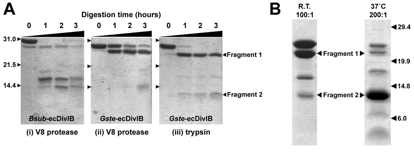
Figure 4.1: (A) Coomassie-stained SDS-PAGE gels illustrating time-course proteolysis of Gste-ecDivIB and Bsub-ecDivIB by trypsin or V8 protease at ambient temperature and a DivIB:protease ratio of 100:1. The mass in kDa and running position of the molecular weight standards is indicated on the left of the gels. Digestion time in hours is indicated above each gel. (B) Coomassie-strained SDS-PAGE gels showing trypsin proteolysis of Gste-ecDivIB using two different temperatures and DivIB:trypsin ratios as indicated above each gel. Fragments 1 and 2 are labeled and where sequenced by Edman degradation.
sample even prior to protease addition (figure 4.1A, panel I, lane 1), suggesting that the N- and/or C-terminus of the recombinant protein is highly susceptible to proteolysis. This truncated fragment develops even further with time, even when the protein is stored at 4º C. At longer incubations times of protein with protease, proteolysis of both Gste-ecDivIB (figure 4.1A, panels II and III) and Bsub-ecDivIB (figure 4.1A, panel I) produced a fragment with an apparent mass of ~ 14 kDa. Irrespective of protease used or origin of species for DivIB, similar proteolysis profiles developed, indicating that DivIB has a consistent domain architecture. Consequently, Gste-ecDivIB was focused on in subsequent experiments designed to identify the major proteolytic fragments.
The time-course proteolysis in figure 4.1A prepared insufficient amounts of protein fragments to allow full identification of the fragments along with any N- or C-terminal cleavage sites. Consequently, several conditions were tried in order to stably and reliably produce sufficient amounts of these fragments for further analysis. Figure 4.1B shows the fragments obtained when Gste-ecDivIB was proteolyzed with trypsin for 3 h using a DivIB:protease ratio of 100:1 at ambient temperature (left panel) or using a DivIB:protease ratio of 200:1 at 37º C (right panel). These two conditions produced large amounts of fragments 1 and 2, respectively, which permitted analysis by mass spectrometry and N-terminal sequencing. Five cycles of N-terminal sequencing indicated that fragment 1 had an intact N-terminus (i.e., G47SPLG), while the N-terminus of fragment 2 (E117WRRI) corresponded to a trypsin cleavage after Arg116. Based on both these N-terminal sequences, mass spectrometry indicated that the C-terminus of both fragments was likely to be Arg230
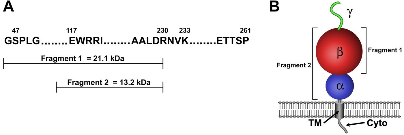
Figure 4.2: (A) Summary of the identity of the two stable proteolytic fragments of Gste-ecDivIB as determined by N-terminal sequencing and mass spectrometry. (B) Illustration of the domain architecture of DivIB, showing the two fragments that were resistant to trypsin proteolysis. The three domains, , and are labeled. Also marked is the transmembrane (TM) domain and the cytoplasmic (CYTO) domain.
(see figure 4.2A). Erring on the side of caution, ensuing sub-cloning of these fragments was extended in the C-terminus to Lys233. These results indicate that the extracytoplasmic region of DivIB comprises of three structurally discrete domains that have here been designated (residues 47-116, 8.0 kDa; blue), (residues 117-230, 13.2 kDa; red), and (residues 231-261, 3.0 kDa; green) (figure 4.2B). Fragment 1 corresponds to +, while fragment 2 corresponds to the isolated domain. The rapid loss of the domain during proteolysis and the spontaneous breakdown of this region even at 4º C indicates that this region is unstructured in the absence of other divisomal proteins. The persistence of fragment 1 for some time during proteolysis suggests that the domain is properly folded when placed in the context of the domain and does not require other divisomal proteins to stabilize its fold. The persistence of the domain for several hours during trypsin proteolysis suggests that it is structurally autonomous in the absence of the domain, the domain and other divisomal proteins.
Amazingly, the domain that was experimentally elucidated here corresponds almost exactly to the polypeptide-transport-associated (POTRA) domain that has been previously predicted, based on bioinformatics analysis, to be present in DivIB/FtsQ proteins and in a class of -barrel outer-membrane proteins involved in the transport of polypeptides (Sanchez-Pulido, Devos et al. 2003). The implications of this are discussed further in section 4.6
4.3. Screening of domains for structure determination
The unstructured nature of the domain in Gste-ecDivIB presents several problems for determining the structure of the entire extracytoplasmic region. Firstly, random coil regions of proteins give disproportionately large signals in NMR experiments due to greater relaxation properties; this combined with regular signal intensities from the structured portion of the protein complicates spectral analysis. Secondly, random-coil regions of proteins after lead to non-ideal behavior of proteins, specifically, non-specific aggregation at high concentration. Indeed, initial screening of the suitability of Gste-ecDivIB by HSQC (see section 3.3.2.3 for a quick discussion of this experiment) showed good dispersion of peaks; an indication that the majority of the protein was folded. However, signal intensity was not uniform and several signals were extremely strong in the middle of the spectrum, an indication that several residues are in a random coil configuration and the protein as a whole was not behaving homogeneously (data not shown).
Sub-cloning of the + domains (Gste-DivIBresidues) produced a stable protein (no visible breakdown after purification, in contrast to Gste-ecDivIB). However, Gste-DivIB upon concentration to ~1 mM readily precipitated. Pprecipitation, however, could be avoided by increasing the ionic strength of the buffer. To maintain a protein concentration of ~1 mM (needed for full structure determination) approximately 700-800 mM NaCl was required as a co-solute. High ionic strength can present a major technical problem during the collection of NMR data. Despite this, a satisfactory HSQC spectrum of Gste-DivIB was collected at ~1 mM (see figure 4.3). Increasing the
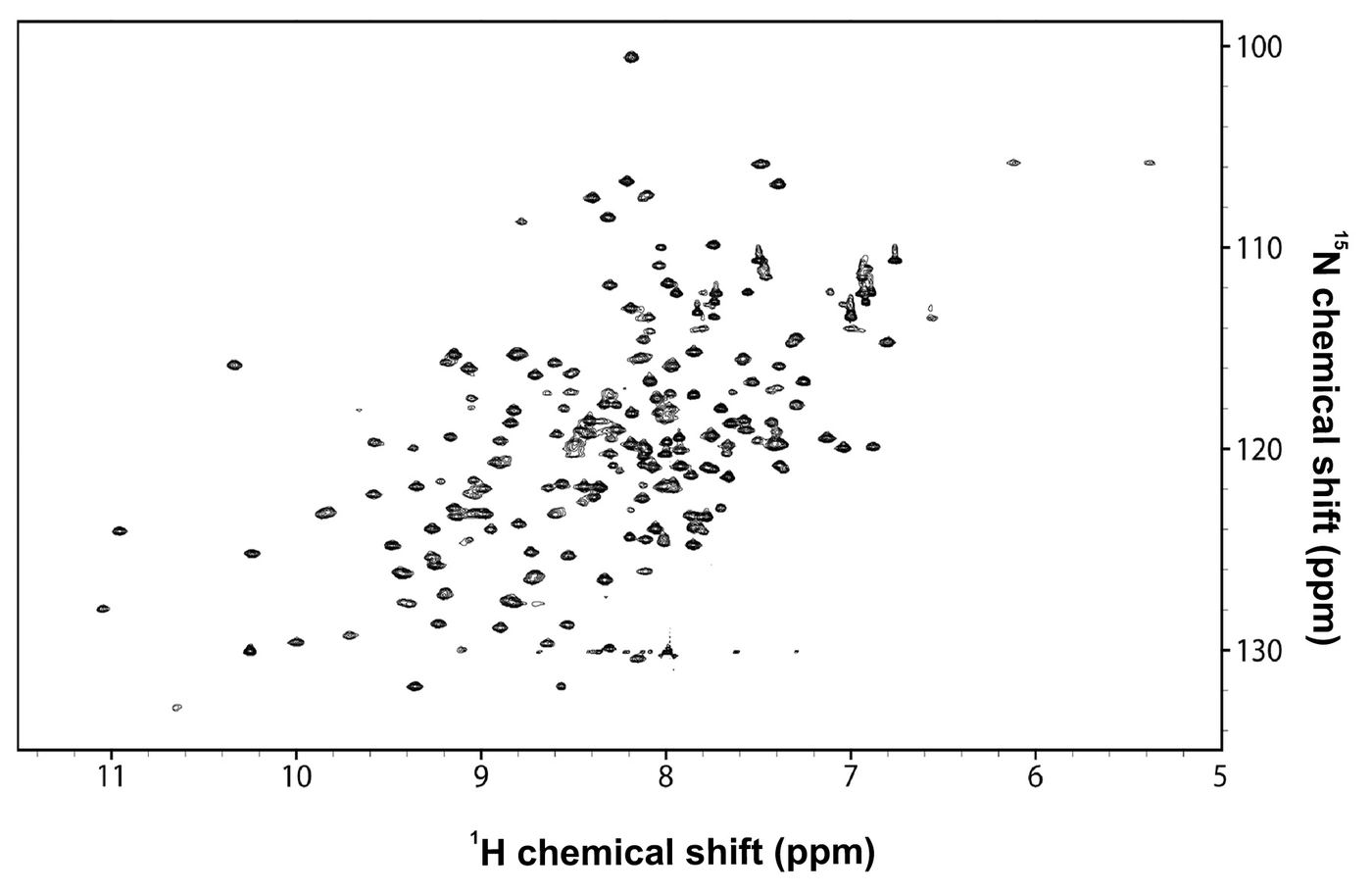
Figure 4.3: 2D 1H-15N HSQC spectrum of Gste-DivIB. Dispersity of peaks in this spectra is quite good in both dimensions indicating that Gste-DivIB is a well folded protein in the absence of the domain.
sample temperature during collection to 35º C significantly increased the homogeneity of signal strengths, highlighting the advantage of using proteins from thermophilic organisms. The dispersion of peaks in figure 4.3 is considerably good, demonstrating that removal of the domain has not caused an unfolding of the protein. The disappearance of strong signals towards the center of the spectrum also indicates that the domain is indeed random coil.
Despite what appears to be a satisfactory HSQC spectrum, a relatively sensitive triple resonance experiment, (the CBCA(CO)NH), failed to generate a suitable spectrum. Triple resonance experiments are necessary for sequence specific chemical shift assignment procedures (discussed below in section 4.4.1). While several assignment strategies are available to make sequence specific assignments, the failure to generate a CBCA(CO)NH spectrum on the instrumentation available at the time suggested full structure determination of Gste-DivIB was intractable. Poor signal strength in the CBCA(CO)NH experiment was likely the result of high salt concentration in the sample combined with the relatively large size of the protein (21.1 kDa) resulting in poor relaxation properties.
Sub-cloning and overexpression of the more proteolytic resistance domain (Gste-DivIBresidues) resulted in a protein fragment with superior solubility properties compared to that of Gste-DivIB. 1 mM Gste-DivIB was readily prepared at pH 6.0 and remained in solution with 20 mM Na3PO4 and 150 mM NaCl. The HSQC spectrum of Gste-DivIB under these buffer conditions at 35º C is given in figure 4.4. Once again, peak dispersion is excellent and peak intensity is highly homogeneous. Gste-DivIB contains 119 residues (included the vestigial N-terminal Gly-Ser from the thrombin cleavage site); four of these are proline residues which do not yield a signal in 1H-15N HSQC spectra due to the absence of a backbone amide proton. Amino acids with amide side chains (Glutamine and Asparagine) yield two additional signals each as these side-chain amides contain two protons covalently bonded to a single nitrogen; these signals lie on a horizontal line in a 1H-15N HSQC spectrum. In addition, the protonated
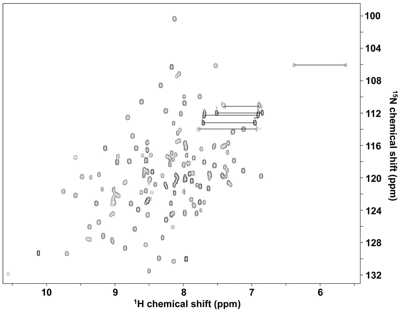
Figure 4.4: 2D 1H-15N HSQC spectrum of Gste-DivIB. Asn and Gln side chain amide protons are marked with horizontal lines. Dispersity of peaks in this spectrum is maintained in both dimensions indicating that Gste-DivIB is well folded in the absence of both and domains. There is approximately 20 peaks too many than expected given the amino acid sequence of Gste-DivIB. There is also some heterogeneity in peak sizes.
N-terminal backbone amide is not expected to yield a signal because of the rapid exchange of these amide protons with solvent water. Thus, figure 4.4 should contain 119 + (2 x 7 amide side chain) – (4 x Pro) – (1 x N-terminal Gly residue) = 128 amide resonances. This calculation significantly underestimates the number of peaks actually present in figure 4.4. The protein was verified to be of the correct mass (by electrospray mass spectrometry) and electrostatically homogeneous (by isoelectric focusing gel); that is, the sample was composed of the correct protein and was not partially post-translationally modified. One possible explanation for the anomalous additional peaks is isomerization between two conformational states. If a certain portion of the protein exists in two differing structural conformations, each conformation would give rise to a unique set of peaks; that is, generating additional peaks than that expected.
Despite the existence of additional peaks in the 1H-15N HSQC spectrum, the most insensitive triple resonance experiment (HNCACB) was successful on Gste-DivIB. The quality of the HNCACB experiment provided enough confidence to pursue full structure determination of the domain, and in addition, determination of the cause of the additional peaks in the 1H-15N HSQC spectrum.
4.4. Structure determination of Gste-DivIB
The determination of protein structures by NMR requires the compilation of a large amount of data from analysis of several different NMR experiments. The sections below describe the process used here to determine the structure Gste-DivIB
4.4.1. Sequence specific assignments.
The first step in NMR structure determination is elucidation of the chemical shifts for all 13C, 15N, and 1H nuclei, a process known as sequence-specific resonance assignment. Complete sequence-specific backbone resonance assignments for Gste-DivIB were obtained from a combination of 3D HNCACO, HNCO, HNHA, HNHB, CBCA(CO)NH and HNCACB NMR experiments. As mentioned above, a successful HNCACB experiment simplifies the sequence-specific assignment procedure. The HNCACB experiment records correlations between the amide 15N and 1H chemical shift of a particular residue and the chemical shift of the C and C resonances of both this residue and the preceding residue. The HNCACB experiment exploits the fact that each amide nitrogen is coupled more strongly to its own C and C than that of the previous residue. As a result, each 1H-15N strip contains intense peaks correlating the backbone amide 1H and 15N resonances of the residue in question with the chemical shifts of its own C and C resonances, and weaker peaks correlating the amide 1H and 15N shifts with the C and C chemical shifts of the previous residue. The C and C correlations are also of opposite sign (typically plotted in different colors or solid lines for positive peaks, broken lines for negative peaks), thus allowing the C and C correlations to be readily distinguished.
Astoundingly, during the assignment procedure for Gste-DivIB, sequence-specific assignments could be determined twice for several regions in the amino acid sequence. That is, sequence-specific assignments were made initially without correlation to the actual amino acid sequence. Upon correlating sequence-specific assignments with amino acids in the sequence, several regions were assigned twice with unique chemical shift assignments. This is highly suggestive that several stretches of amino acids exist in two distinct structural conformers. Figure 4.5 illustrates the HNCACB sequence-specific assignments for residues 223-225 arising from two conformers. It can be seen that despite these signals arising from the same amino acids, there are distinct and, in some cases, extreme differences between conformers in the chemical shifts for the backbone nuclei (C and C sidechain nuclei have resonance frequencies that are more sensitive to the sidechain chemistry rather than backbone structure and do not vary as much). For example, the backbone 15N nuclei of reside 223 has a chemical shift of 131.9 ppm in conformer 1 and a chemical shift of 120.1 ppm in conformer 2. The radical change in chemical shift (almost 12 ppm) between conformers is indicative of a drastic change in the structure, particularly of the backbone in this region. Sequence-specific sidechain assignments for each conformer were obtained from analysis of 3D HC(CO)NH-TOCSY, and C(CO)NH-TOCSY, and HCCH-TOCSY spectra. Despite the confusion that can arise from having two sets of peaks for a subset of amino acids, the 1H, 15N and 13C assignments for each conformer was determined, with the exception of 1HN and 15N of residue Q213, aromatic 13C resonances, and the labile protons of lysine and arginine residues and their associated side-chain 15N. 13C’ assignments were made for all residues.
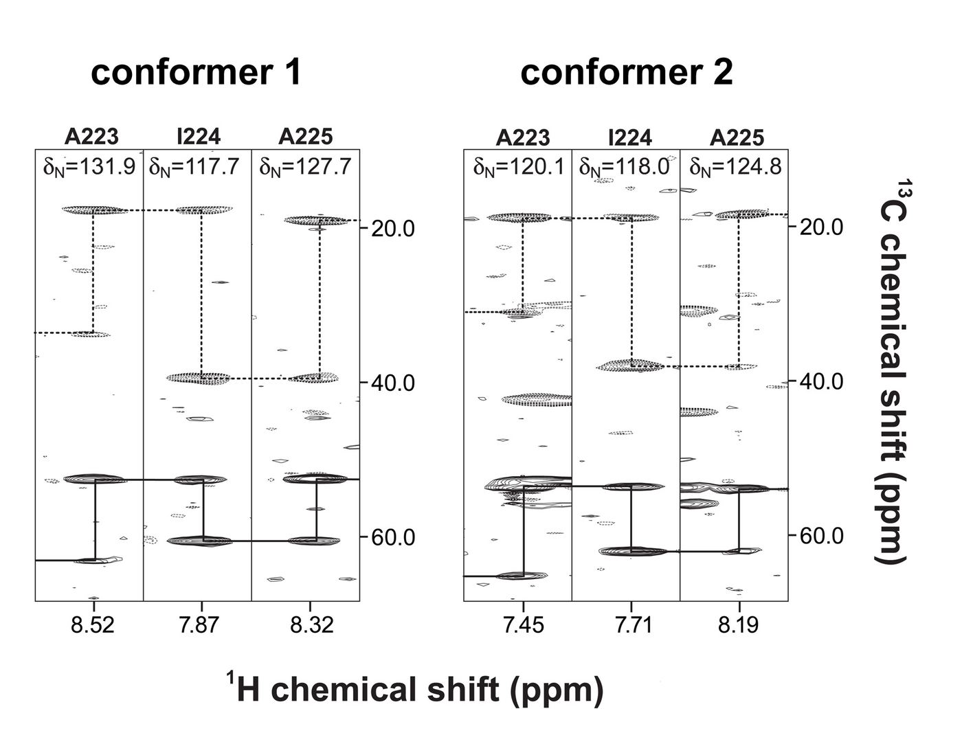
Figure 4.5: Selected 1H-15N strips from a 3D HNCACB spectrum of Gste-DivIB. Backbone amide strips for residues 223-225 for alternate conformers 1 and 2. Each 1H-15N strip contains strong peaks correlating the chemical shift of the backbone amide proton of the residue in question (horizontal axis) with the chemical shift of its own C and C resonances (vertical axis), and weaker peaks correlating the backbone amide proton chemical shift with the chemical shifts of the C and C nuclei of the previous residue in the amino acid sequence. In the figure above, C correlations are depicted in solid lines and C correlations are shown by dashed lines. Note, each conformer has similar C and C shifts for each residue as expected, yet the backbone 1HN and 15N differ greatly between conformers.
4.4.2. The origin of the two distinct structural conformers.
The above assignment procedure successfully determined almost complete backbone and side chain resonances for what appeared to be two distinct structural conformers. The 1H-15N HSQC spectrum of Gste-DivIB with the amide resonances labeled with their amino acid numbers is shown in figure 4.6 (panel A). The predominant conformer is labeled in black with the minor conformer labeled in red. It can be seen that 21 amino acids appear to exist in two different states. Furthermore, they are predominately located in the C-terminal region of the domain.
What could be the cause of two distinct structural states in Gste-DivIB? One possible explanation could be cis-trans isomerization around a peptide bond preceding a Proline residue. An XXX-Pro peptide bond can exist in a cis or trans configuration with little energetic difference between the two, although trans is slightly favored. This is in contrast with other peptide bonds in which the trans configuration is greatly favored over the cis configuration. As a result, cis peptide bonds before proline residues are frequently observed and in some cases, proteins have been noted to exchange between two structural conformers as a result of flipping between a trans and cis conformation. Indeed, this situation is observed in the 1H-15N HSQC spectrum of Gste-DivIBThe peaks that give rise to the minor conformer are only slightly less intense than the major conformer (see the peaks are G204 for an example).
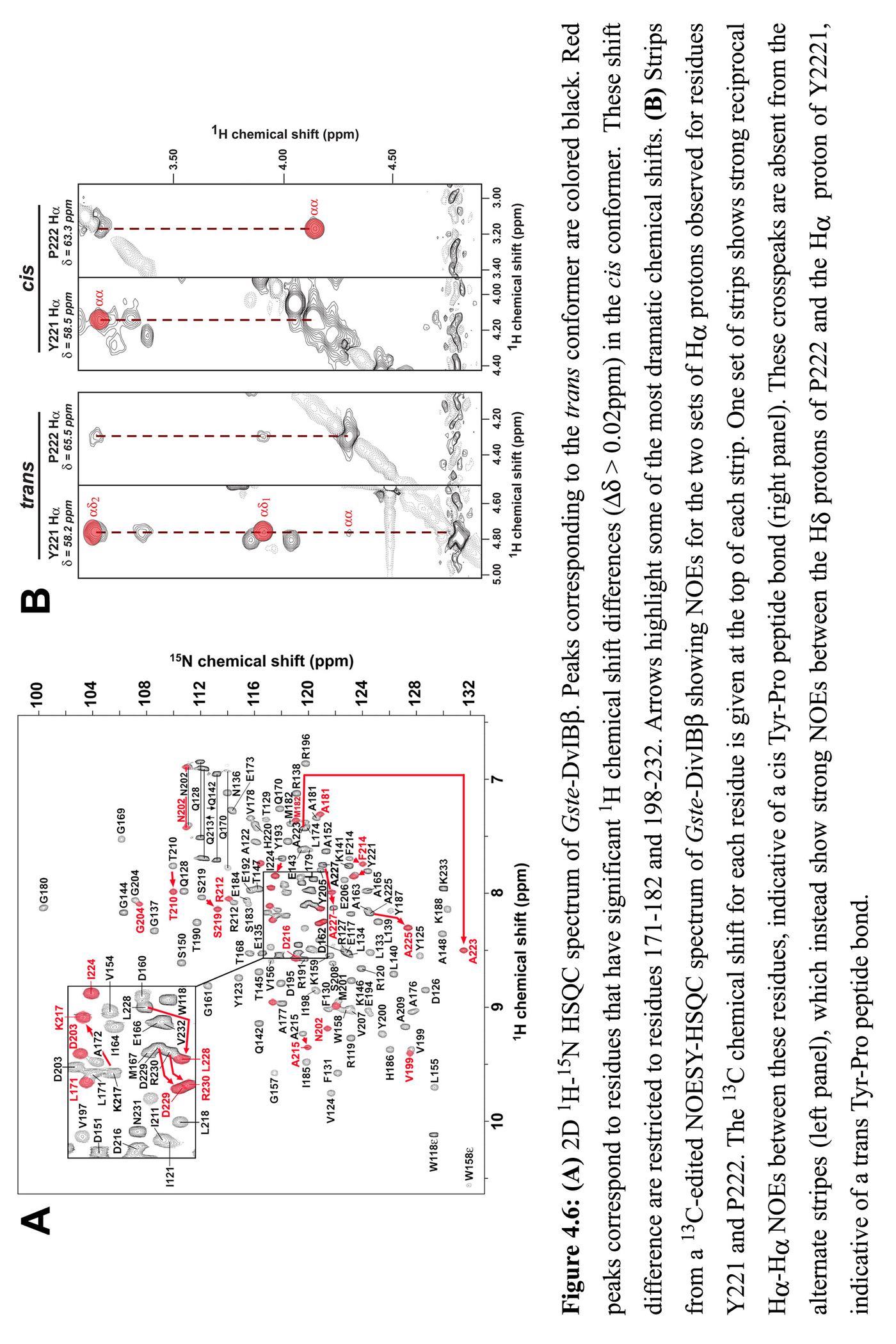
In addition, since there exists a large energetic barrier for exchanging between the cis and trans states, the kinetics of flipping between conformers is slow (on the time scale of seconds). Typically, a pulse sequence for an NMR experiment is on the time scale of microseconds to milliseconds, so data collected during a pulse sequence samples the structural state over that short period. If proline isomerization is causing distinct structural conformers, then it is to be expected that each conformer should be able to be discretely witnessed during a pulse sequence rather than an average of the two states. Indeed, that is what is observed in the 1H-15N HSQC spectrum of Gste-DivIB
More conclusive evidence for cis-trans proline isomerization can be seen in panel B of figure 4.6. Strips from a 13C-edited 3D NOESY-HSQC spectrum of Gste-DivIB shows NOEs for the two sets (the two conformers) of H protons observed for residues Y221 and P222. NOE experiments are designed to show the proximity of protons to one another. One set of strips shows strong reciprocal H-H NOEs between Y221 and P222, indicative of a cis Tyr-Pro peptide bond (right panel). These crosspeaks are absent from the alternate strips (left panel), which instead show strong NOEs between the H protons of P222 and the H proton of Y221, indicative of a trans Tyr-Pro peptide bond. Thus, it is concluded that Gste-DivIB exists in two structural conformations that result from cis-trans isomerization about the Y221-P222 peptide bond.
4.4.3. Generation of restraints for structure determination
In order to calculate structures from NMR data, three types of experimental data are used as restraints during the calculations. The dihedral backbone and angles are determined from measured chemical shifts of backbone nuclei, distance constraints between protons are calculated and finally hydrogen bonds are declared based on preliminary calculated structures in combination with deuterium-exchange experiments.
4.4.3.1 Angle Restraints
Dihedral angles were predicted using the TALOS program (Cornilescu, Delaglio et al. 1999). TALOS allows empirical prediction of a protein’s backbone and using a combination of five kinds of chemical shift assignments (i.e., 1H, 13C, 13C, 13CO, 15N) determined above. This approach is based on the empirical observation that differences between chemical shifts determined from folded proteins and their corresponding random coil values are highly correlated with protein secondary structure. TALOS attempts to match chemical shift assignments with values from a database of known structures. Automated analysis of the chemical shifts of Gste-DivIB using TALOS generated xx and xx dihedral-angle restraints for the trans conformer and xx and xx dihedral-angle restraints for the cis conformer.
In addition to and angles, sidechain 1 angles were also estimated where possible. The 3D HNHB experiment provides correlations between the backbone amide proton (HN) of a residue and its sidechain H protons. The intensity of each crosspeak is related to the size of the 3JHN-H coupling, which in turn is related to the sidechain 1 angle. Thus, analysis of the 3JHN-H couplings observed in an HNHB experiment in combination with the H-H and HN-H NOE intensities observed in 15N-edited and 13C-edited NOESY experiments (which are also 1-dependent) can provide an estimate of the sidechain 1 angle as well as stereospecific assignments for the pair of -methylene protons. Using this approach, stereospecific assignments for 18 pairs of H protons was determined as well as estimates for 29 1 angles for both cis and trans conformers.
4.4.3.2 Distance constraints
Analysis of the 3D 15N- and 13C-edited NOESY-HSQC spectra of Gste-DivIB reveal dipolar correlations between spatially proximal hydrogen atoms. Dipolar correlations (crosspeaks) are typically observed for pairs of hydrogen atoms that are separated by less than 5.5 Å, with the intensity (I) of the crosspeak (for immobile protons) inversely proportional to the sixth power of the internuclear distance, r (i.e., I 1/r6). Thus, each crosspeak volume provides an estimate of the distance between the two correlated protons. Analysis of 3D 15N- and 13C-edited NOESY-HSQC spectra is a complicated procedure, which is in part conducted automatically by computer software. The sequence specific assignments generated above for each conformer were mapped onto the NOESY spectra. These assignments are used as a basis for finding the dipolar correlations from other proximal protons in the spectrum. The details of this process are discussed further in section 4.4.4.
4.4.3.3 Hydrogen bonds
Hydrogen bonds were determined by rapidly exchanging Gste-DivIB into D2O (see section 2.x.x for details) and monitoring for the disappearance of amide proton signals in the 1H-15N HSQC experiment. As the HN protons of proteins are labile, they rapidly exchange with solvent protons, or when D2O is used as a solvent, with deuterium. Deuterium does not give a signal in a 1H-15N HSQC experiment. When an HN participates in a hydrogen bond, it exchanges with solvent at a much slower rate, hence hydrogen bonded HN protons can remain in hydrogen bonds for tens of minutes to days and continue to give rise to a signal in a 1H-15N HSQC experiment (a deuterium exchange experiment). Collection of a 1H-15N HSQC spectra 30 minutes to an hour after exchange into D2O will result in hydrogen bonded HN protons still being visible. The electro-negative nuclei (typically oxygen) the HN proton is hydrogen bonded to can not be directly determined this way, however, based on preliminary structures of sufficient accuracy, these hydrogen bond partners can be predicted. These predicted hydrogen bonds are utilized in final structure calculations and greatly increase the precision and accuracy of the final structures. Analysis of a deuterium exchange experiment on Gste-DivIB after 30 minutes of exchange into D2O, in conjunction with preliminary structure calculations, revealed the presence of 44 hydrogen bonds which were used in final structure calculations.
4.4.4 Structure calculations
Until recently, NOESY spectra were typically assigned manually and iteratively. That is, the sequence-specific resonance assignments would first be used to assign unambiguous correlations in the NOESY spectra (discussed briefly in section 4.4.3.2.). Preliminary structures would then be calculated, and then new assignments would be made based on the preliminary structures. This process would be iterated until as many NOESY peaks as possible were assigned.
During recent years, a number of computer programs have been introduced that allow NOESY spectra to be automatically assigned using an iterative procedure of assignment, calculation, and re-assignment. This process superficially resembles the manual approach but is far more time efficient; so long as sequence-specific resonance assignments are >90% complete, the program used in this laboratory (CANDID) typically assigns 85–90% of all NOESY peaks and produces structures that are as good or better than that obtained by manual analysis.
Thus, the crosspeaks in the 3D 15N- and 13C-edited NOESY-HSQC spectra of Gste-DivIB were integrated and the list of chemical shifts and intensities of each peak were used as input to the program CANDID (Herrmann, Guntert et al. 2002; Herrmann, Guntert et al. 2002; Jee and Guntert 2003), along with a complete list of the sequence-specific resonance assignments. Care was taken to carefully separate cis assignments from trans assignments and calculations with data for each conformer run individually. Other input data included restraints for the experimentally observed hydrogen bonds (Section 4.4.3.3), the and backbone dihedral-angle restraints derived from TALOS (Section 4.4.3.1), the 1 angles derived from analysis of HNHB spectra (Section 4.4.3.1), and the declaration of 18 H stereospecific assignments (Section 4.4.3.1).
CANDID was then used to automatically assign the spectra and calculate structures of Gste-DivIB in both cis and trans conformers, using seven iterative cycles of spectral assignment and structure calculation. This process took approximately 45 minutes per conformer running on a cluster of 10 Apple dual-processor Xserves. This led to a total of 2746 (trans) and 2544 (cis) meaningful interproton distance restraints, which can be divided into 363 (trans) and 324 (cis) intraresidue, 618 (trans) and 556 (cis) sequential, 632 (trans) and 579 (cis) medium range (|i–j| 5), and 1133 (trans) and 1085 (cis) long range (|i–j| < 5) restraints (table 4.1). These restraints were used in combination with the dihedral-angle and hydrogen-bond restraints to calculate 600 structures using CYANA and the best 60 (chosen on the basis of lowest penalty function values) were then refined via dynamical simulated annealing using X-PLOR (Weis, Brunger et al. 1990). The 25 structures with lowest molecular energies and no restraint violations were selected as representative of the Gste-DivIB structure (results summarized in table 4.1). Each conformer was submitted to the protein databank (http://www.rcsb.org/pdb/) and designated a PDB ID; 1YR1 for the trans conformer and 2ALJ for the cis conformer.
Table 4.1: Structural statistics for the ensembles of cis and trans Gste-DivIB structures
trans (1YR1) cis (2ALJ)
Experimental restraints1
Interproton distance restraints
Intraresidue 363 324
Sequential 618 556
Medium range (i–j 5) 632 579
Long range (i–j 5) 1133 1085
Hydrogen-bond restraints2 88 88
Dihedral angle restraints 200 197
Total number of restraints per residue 25.5 23.8
Mean r.m.s. deviations from experimental restraints3
NOE distances (Å) 0.01509 ± 0.00027 0.01278 ± 0.00034
Dihedral angles (deg.) 0.277 ± 0.015 0.340 ± 0.024
Mean r.m.s deviations from idealized geometry4
Bonds (Å) 0.00216 ± 0.00004 0.00214 ± 0.00003
Angles (deg.) 0.340 ± 0.005 0.336 ± 0.002
Impropers (deg.) 0.226 ± 0.004 0.202 ± 0.005
Mean X-PLOR energies (kcal mol-1)
ENOE5 31.3 ± 1.1 21.5 ± 1.2
Ecdih5 0.94 ± 0.10 1.39 ± 0.20
Ebond 8.79 ± 0.35 8.67 ± 0.27
Eimproper 7.75 ± 0.29 6.21 ± 0.32
Eangle 60.2 ± 1.6 58.7 ± 0.7
Erepel 41.8 ± 1.5 38.9 ± 1.4
R.m.s. deviation to mean coordinate structure (Å)6
Backbone atoms 0.17 ± 0.03 0.18 ± 0.02
All heavy atoms 0.66 ± 0.04 0.64 ± 0.04
Residues in most favored Ramachandran region 85% 80%
1Only structurally relevant restraints, as defined by CYANA, are included.
2Two restraints were used per hydrogen bond.
3All statistics are given as mean ± S.D.
4Idealized geometry is defined by the CHARMM force field as implemented in X-PLOR.
5Final values of the square-well NOE and dihedral-angle potentials were calculated with force constants of 50 and 200 kcal mol–1 Å–2, respectively.
6r.m.s.d. values were calculated over the well-defined regions of the structure (residues 118–229 for the trans isomer and residues 118–223 for the cis isomer).
4.5 The 3D structure of Gste-DivIB
The solution structures of the trans and cis conformers of Gste-DivIB are shown in figure 4.7(panels A–C) and figure 4.7(panels D–E), respectively. Panels A and D show a stereoview of an overlay of 25 Gste-DivIB structures for the trans and cis conformers respectively. In each case, the backbone is well structured for the majority of the protein. Only in the cis conformer is there a significant amount of unstructured backbone (the last ten residues). The backbone r.m.s.d over the structured region is 0.17 Å ± 0.03 Å for trans (residues 118-229) and 0.18 Å ± 0.02 Å for the cis conformer (residues 118-223). All heavy atom r.m.s.d over the same range is 0.66 Å ± 0.04 Å and 0.64 Å ± 0.04 Å for trans and cis respectively. The dihedral angles over the most structured regions comply with the most favored regions of the Ramachandran plot with 85% and 80 % of dihedral angles falling in the most favored region for trans and cis respectively.
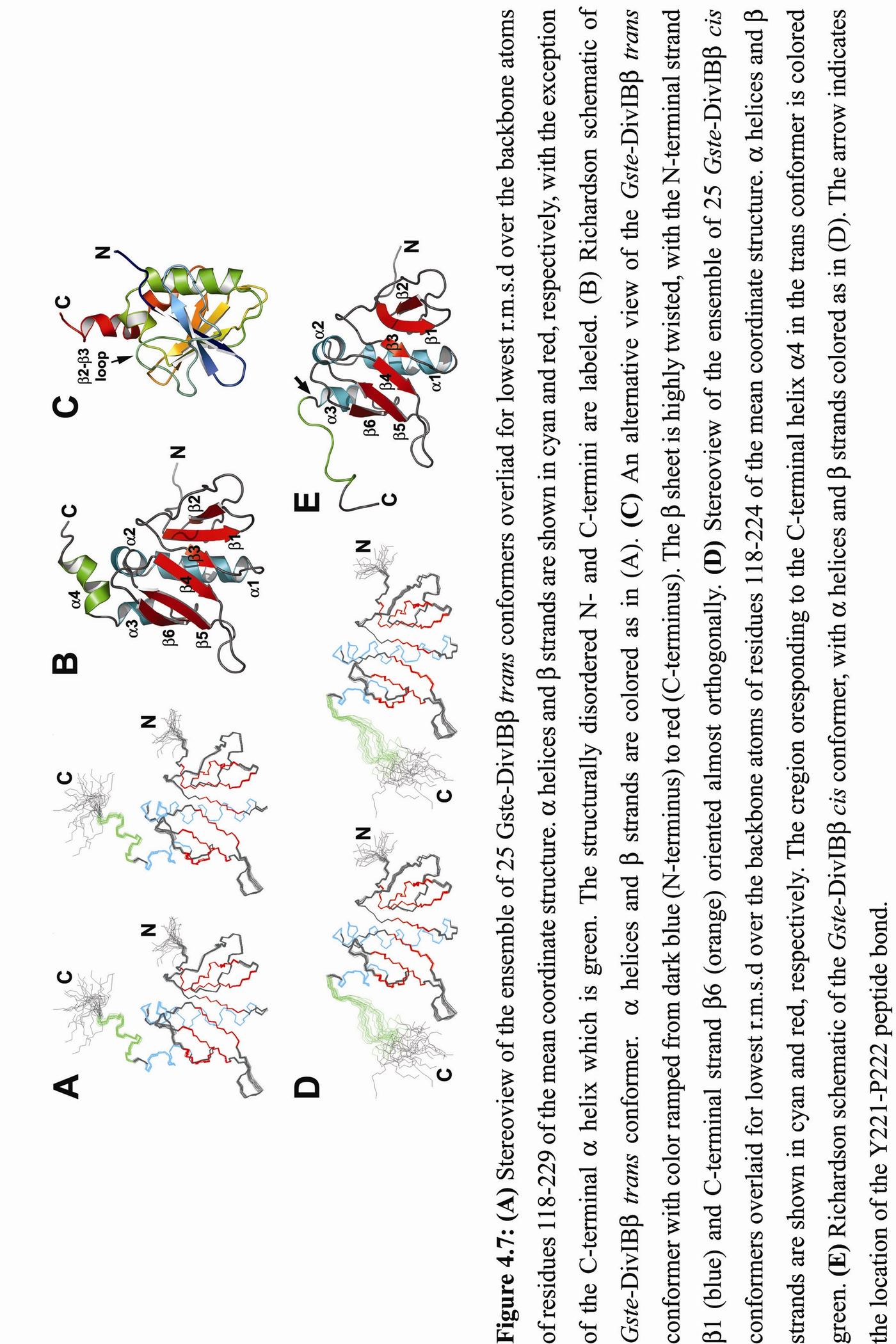
The structure of the trans isomer of Gste-DivIB, which represents ~60% of the sample population under the chosen experimental conditions, comprises a six-stranded mixed sheet flanked on one side by three helices. A fourth helix (green in Fig. 4.7A,B) is located C-terminal to the Y221–P222 isomerization site. The sheet is highly twisted, with the first and last pair of strands oriented almost orthogonally (Fig. 4.7C). The structure contains a rare left-handed crossover connection between parallel strands 3 and 4 (Fig. 4A–C). This connection includes helices 1 and 2, which are positioned parallel and perpendicular to 4, respectively. More than 99% of connections between parallel strands in -- motifs are right-handed. In rare instances where left-handed
connections do occur, they are often functionally important, as in subtilisin and asparaginase (Miller, Rao et al. 1993). This suggests a possible functional role for the 3-1-2-4 region of DivIB.
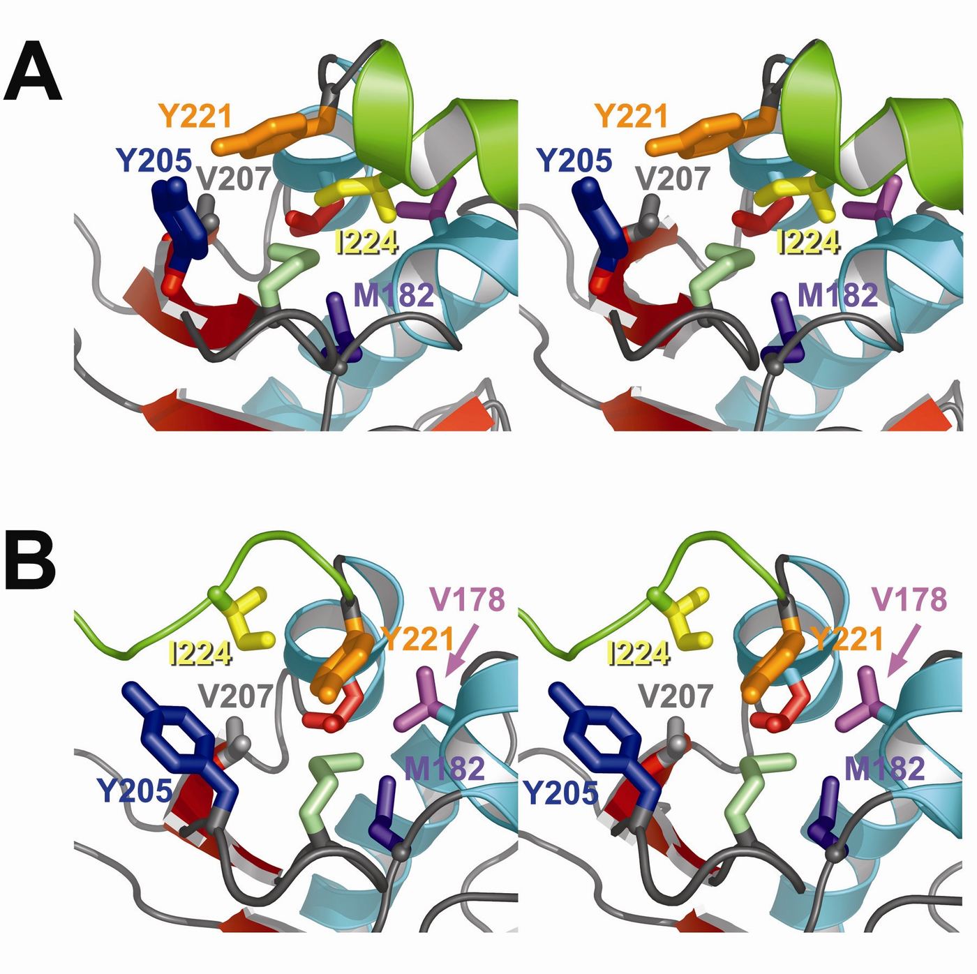
The structures of the cis and trans conformers of Gste-DivIB are very similar except for the C-terminal 11 residues; the mean structures can be superimposed over residues W118–L218 with a backbone r.m.s.d of 0.8 Å. However, the presence of the cis Y221–P222 peptide bond causes an abrupt reversal in the polypeptide chain direction centered around P222 and the C-terminal 4 helix found in the trans conformer is replaced by a more extended and less well defined structural element in the cis conformer (Fig. 4.7D,E). The reversal of the polypeptide chain direction in the cis conformer causes a dramatic rearrangement of buried hydrophobic residues near the isomerization site. In the cis conformer (compare figures 4.8A and B), the sidechain of Ile224 switches location to the opposite side of the Tyr221 aromatic ring, which rotates towards 2 to a position above the sidechain of Met201. This repositioning of Tyr221 creates room for a marked reorientation of the sidechain of Tyr205; in the trans isomer the aromatic ring of Tyr205 is completely solvent exposed whereas it makes more intimate contact with the core of the domain in the cis conformer. The space created by relocation of Ile224 is filled by slight reorientations of the sidechains of Met201 and Val178 in the cis isomer.
Figure 4.8: Stereoview of the C-terminal hydrophobic cores of (A) trans and (B) cis conformers of Gste-DivIB. Helices and strands are colored as in fiugre 4.7. Sidechains of buried hydrophobic residues are drawn as colored tubes. Residues M201 (sage) and L218 (red) are not labeled for the sake of clarity. The sidechain of Y205 is not part of the hydrophobic core, but it adopts a markedly different and less solvent-exposed orientation in the cis isomer.
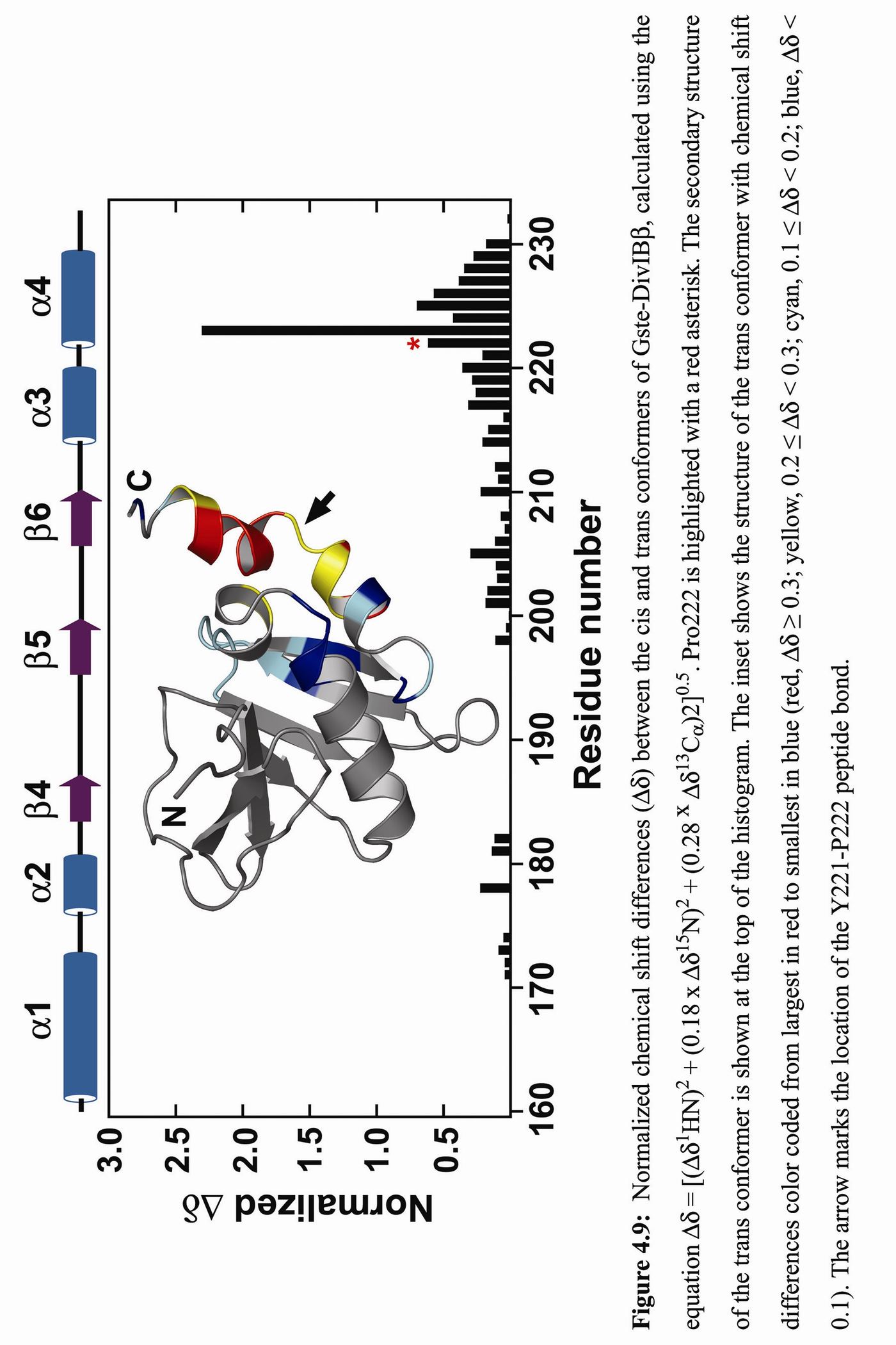
All of the residues that show chemical shift variations between the cis and trans conformers are spatially proximal to the Tyr221–Pro222 isomerization site (figure 4.9), with the largest differences due to altered ring current shifts caused by reorientation of the sidechains of Tyr205 and Tyr221.
The structural variation between the cis and trans conformers of Gste-DivIB is more dramatic than has been previously observed for other similarly sized proteins that undergo proline cis-trans isomerization, such as the Itk SH2 domain, the viral coat protein of bacteriophage MS2, and the TB domain of human fibrillin-1 (Andreotti 2003). Moreover, in striking contrast with these proteins, it is the cis rather than the trans isomer that presents the most extended, solvent-exposed conformation.
A search of the Protein Data bank using DALI (Holm and Sander 1993) and Combinatorial Extension (Shindyalov and Bourne 1998) returned no meaningful structural homologs of Gste-DivIB. This was not entirely surprising as BLAST searches with DivIB and FtsQ sequences consistently return orthologs of these proteins but no other closely related sequences. Thus, the Gste-DivIB structure is the first example of this unusual mixed / architecture.
4.6 Discussion and Conclusions
The extracytoplasmic region of DivIB/FtsQ is necessary and sufficient for efficient cell division to take place (Chen, Weiss et al. 1999; Katis and Wake 1999). However, prior to this study, nothing was known about the molecular architecture of this key functional region. Here, it has been shown that the extracytoplasmic domain of DivIB comprises three domains, designated, from N- to C-terminus, , and . The domain appeared to be unstructured in the absence of other divisomal proteins while both the and domains are structurally autonomous. Based on primary structure homology across the DivIB/FtsQ family, the domain architecture is expected to ubiquitous.
The domain elucidated in this study is almost exactly co-incident with the POTRA domain, an, until now, theoretical domain predicted on the basis of bioinformatic analysis alone. The POTRA domain is present in the Toc75 subunit of the chloroplast import mechanism (Jarvis and Soll 2001), the Omp85/D15 protein family that play a role in assembly of outer-membrane proteins (Voulhoux, Bos et al. 2003) and in Serratia marcescens hemolysin IB-related proteins that reside in the outer-membrane of Gram negative bacteria and mediate export of virulence factors (Hertle 2000). The original proponents of the POTRA domain suggested that this domain may function as a chaperone for secreted or imported, unfolded polypeptides (Sanchez-Pulido, Devos et al. 2003). Therefore, it is possible that the domain of DivIB itself can act as a chaperone for unfolded proteins in the divisome. One obvious candidate is the FtsL protein which is unfolded and unstable in the absence of other divisomal proteins (see chapter 3) and FtsL is rapidly degraded in the absence of DivIB at the non-permissive temperature (Daniel and Errington 2000). Alternatively, the domain may act as an intramolecular chaperone for the unstructured domain. Such a role for the domain is particularly highlighted given the structure of the domain. Figure 4.7 (panels A and C) draw attention to the close proximity of the N- and C-termini when in the trans configuration. In the cis configuration, the N- and C-termini point away form each other, suggesting intramolecular chaperone activity would be accompanied by the trans configuration state only.
Because of their essential role in cytokinesis, divisomal proteins are considered potential antibacterial targets (Margalit, Romberg et al. 2004). A possible complication with this approach, however, is that, with the exception of ZapA, there are eukaryotic structural homologs of all bacterial cell division proteins whose structures have been determined, including tubulin (FtsZ), actin (FtsA), and the ubiquitous RNA-binding motif (ZipA and FtsN). In contrast, DivIB is the first example of a membrane-tethered divisomal protein for which there is not a structural counterpart in eukaryotes. Thus, in organisms where they are essential, DivIB and FtsQ might be good targets for therapeutic intervention. It is expected that the elucidation of the structure of the domain of DivIB is an important step in this direction.